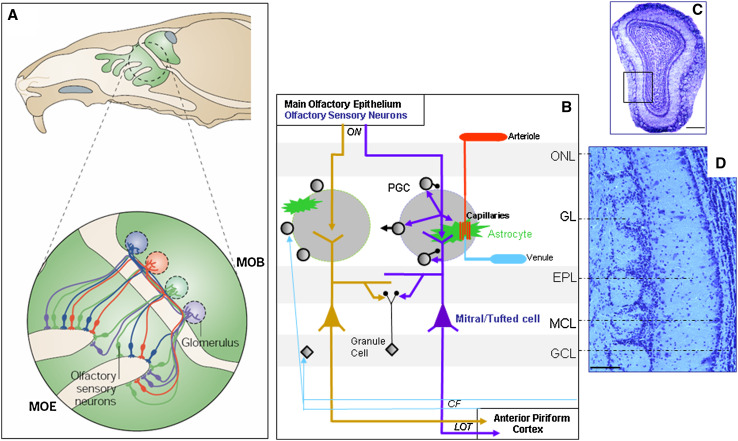
Fig. 1.
The olfactory bulb, first structure for olfactory information processing in the brain. a Top: Sagittal view of a rodent head showing in green the location of the MOE at the very end of the nasal cavity and the MOB at the anterior position in the brain. Bottom: OSNs, primary receptor cells located in the MOE, which express the same odorant receptor, converge onto the same glomeruli in the MOB. b Olfactory glomeruli, the round-shaped neuropils located at the surface of the olfactory bulb, about 100 μm deep just under the olfactory nerve layer (ONL), are constituted by incoming convergent OSN axon fibers. In each glomerulus, projections of OSNs ramify and constitute synapses (purple arrows) with the apical dendritic tufts of principal excitatory cells (M/TCs) and inhibitory interneurons (periglomerular cells, PGCs). M/TCs and PGCs interact via dendrodendritic synapses: PGC dendrites project inhibitory GABAergic synapses onto M/TC dendrites (lower black circle), whereas M/TC dendrites make glutamatergic contacts onto PGC dendrites (purple arrow). PGCs also make GABAergic synapses onto OSN axonal terminals (upper black circle). This interneuronal population includes short axon cells which could allow lateral interactions between distant glomeruli (black arrow to the left). Numerous astrocytic projections are also part of this neuropil that they compartmentalize: they enwrap both synapses and blood vessels (green star-like branches) cleaning up glutamate and regulating local hemodynamics. Note that a very dense and complex vascular network is present at the glomerular level (arteriole, capillaries, venule). The dendrodendritic interactions in the external plexiform layer (EPL) allowing lateral inhibition between M/TCs are due to the activation of local inhibitory interneurons, the granule cells (GCs). Each M/TC is specific to one glomerulus and sends information via the lateral olfactory tract (LOT) mainly to the anterior piriform cortex and to the limbic system without a thalamic relay. These structures send back centrifugal fibers (CF) to the MOB for feedback regulation. c Coronal section of a mouse MOB (top dorsal, bottom ventral, left medial, right lateral). Inset lower magnification of image d. Scale bar 500 μm. d Anatomical view of MOB layers. Note the olfactory glomeruli aligned close to the MOB surface and the very thin layer of M/TCs. Scale bar 100 μm. GL glomerular layer, GCL granule cell layer, MCL mitral cell layer, ON olfactory nerve. (a Adapted from reference [1] with permission from Macmillan Publishers; copyright 2004)

