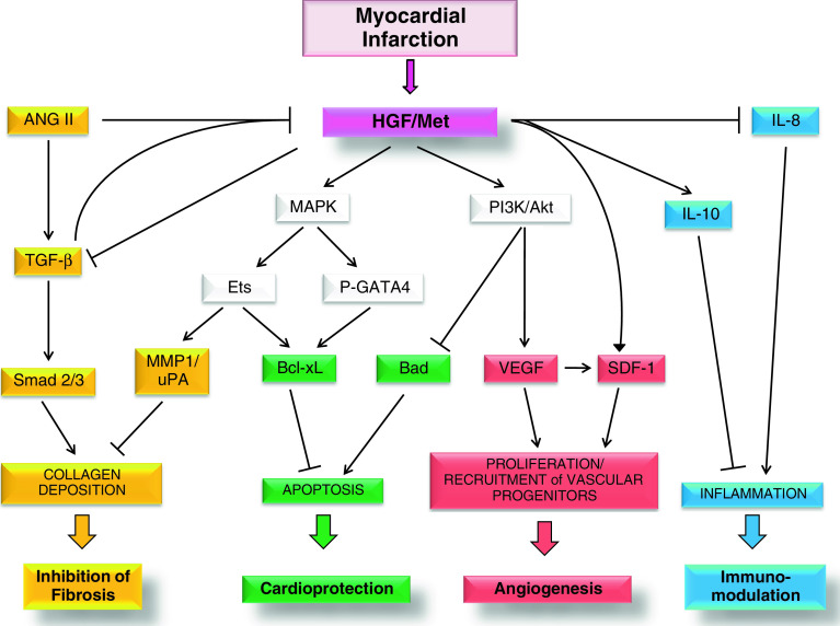
Fig. 1.
HGF and Met receptor levels increase after MI. Activation of Met receptor at the surface membrane of cardiomyocytes and interstitial cells leads to stimulation of different signalling pathways and biological responses. The local increase in secreted HGF results in inhibition of TGF-β signalling in myofibroblasts and ultimately in the inhibition of collagen deposition and fibrosis (yellow). Conversely, TGF-β and ANG II, another profibrotic factor, behave as negative regulators of HGF local production. Stimulation of Met receptor results in MAPK-mediated phosphorylation of GATA4 and Ets, which stimulate the antiapoptotic activity of Bcl-xL, while the PI3K/Akt axis inhibits Bad, a proapoptotic factor. Thus, activation of Met downstream signalling protects cardiomyocytes from programmed cell death (green). The Ets transcription factor also acts in the prevention of fibrosis by inducing extracellular matrix remodelling through MMP1 and uPA (yellow). Proliferation and recruitment of vascular progenitors are also promoted by Met activation due to induction of VEGF and SDF-1, finally enhancing angiogenesis (red). Activation of Met receptor in immune cells reduces inflammation (blue). See text for more details. ( activation,
  inhibition)

