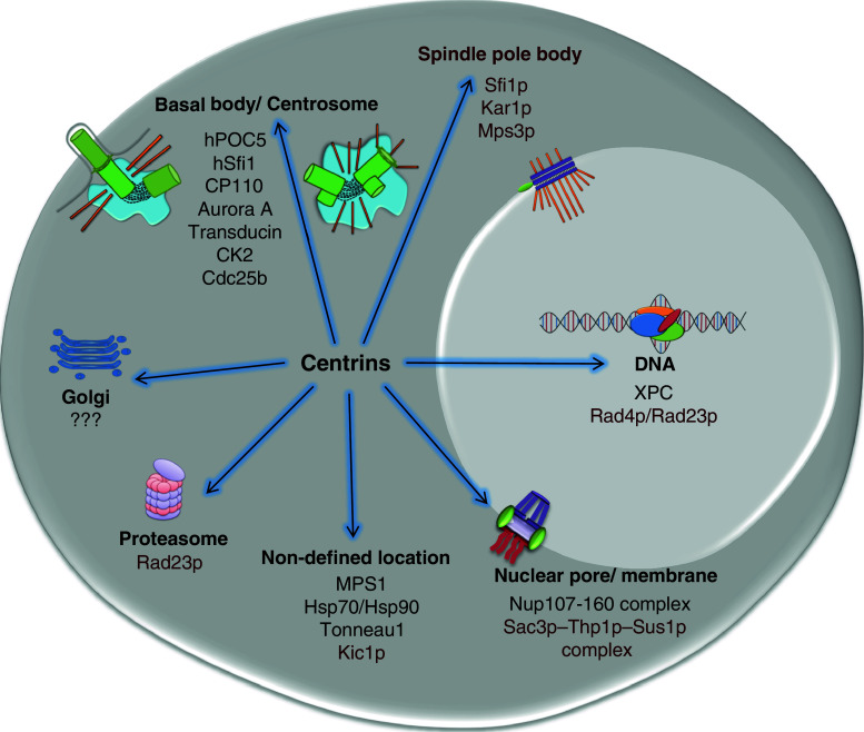
Fig. 1.
The localisation and interactions of centrins. Cartoon shows centrin interactions in higher eukaryotes and in yeast (in brown), indicating the organelles or structures where centrins were found to associate with their respective partners, as discussed in the text. The references describing these interactions are as follow: hSfi1 [221], hPOC5 [219], CP110 [174], Cdc25b [76], XPC [105], Aurora A [145], MPS1 [115], CK2 [222], Transducin [147], Nup107-160 complex [108], Hsp70/Hsp90 [100], Kic1p [153], Tonneau1 [223], Rad4 [106], Rad23p [106], Sfi1p [142], Kar1p [151], Mps3p [152], Sac3p–Thp1p–Sus1p complex [107], golgi localisation [160]. Note that the elements of the idealised cell shown here are not to scale

