Fig. 5.
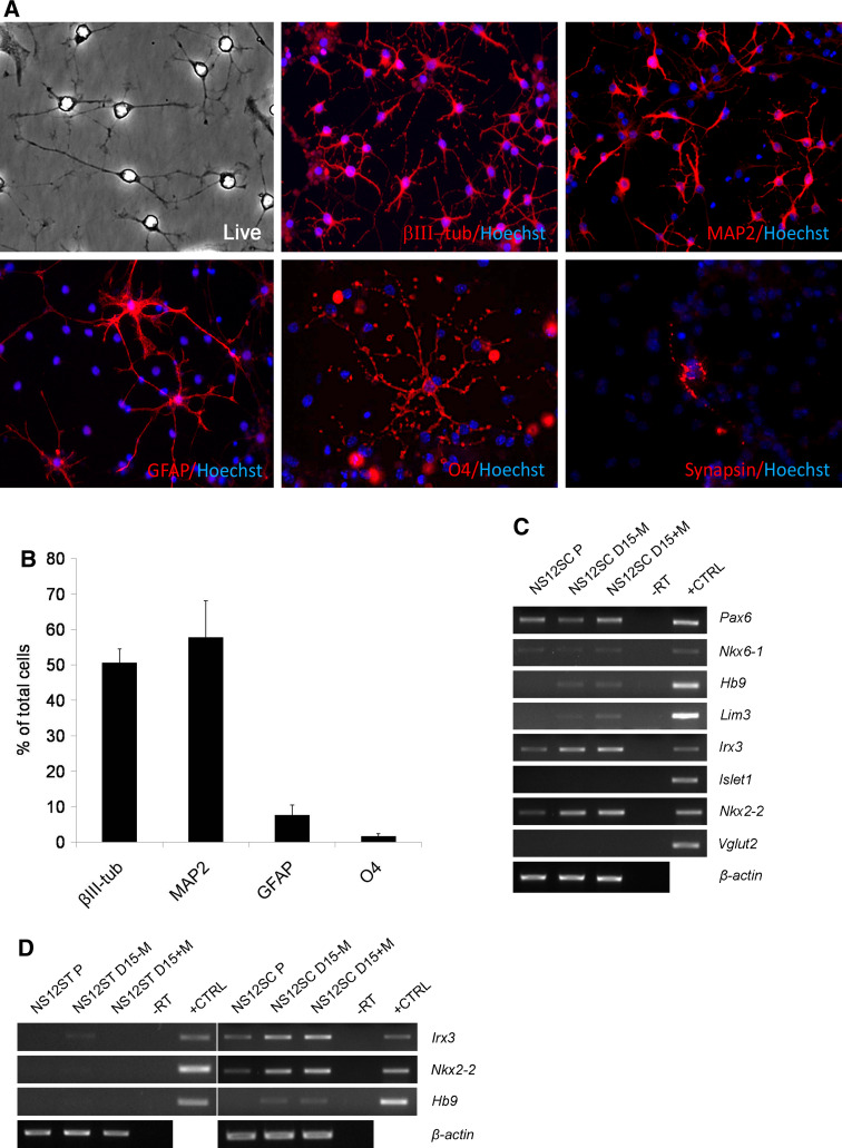
Neuronal differentiation of NS12SC cells. a After 23 days of differentiation in vitro, NS12SC cells gave rise to βIII-tubulin-, MAP2-, GFAP-, and O4-positive cells. A representative synapsin-positive cell is shown after 21 days of differentiation (×4, magnified after acquisition). b At the end of differentiation, 50.5 ± 3.9% of cells were immunopositive for βIII-tubulin, 57.7 ± 10.3% for MAP2, 7.5 ± 2.8% for GFAP, and 1.5 ± 0.7% for O4 (n = 1,068 cells for βIII-tubulin; n = 1,184 cells for MAP2 and GFAP; n = 1,645 cells for O4) (columns represent averages, error bars standard deviations). c Gene expression analysis by RT-PCR on NS12SC cells in proliferation (P) and after 15 days differentiation in the absence (D15-M) or in the presence (D15+M) of morphogens. d Spinal marker expression was restricted in NS12SC and not detectable in NS12ST cells. Mouse fetal brain was used as positive control (+CTRL); -RT was the negative control

