Fig. 1.
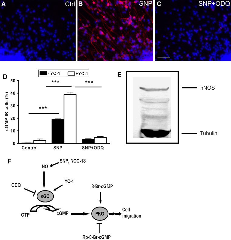
Functional NO/cGMP signaling in early developing human nerve cells. The presence of NO-sensitive sGC was demonstrated by immunocytochemical detection of cGMP. The hNPCs were allowed to migrate and differentiate on poly-d-lysine/laminin-coated substrates for 72 h. Cultures were exposed for 20 min to a 1 mM IBMX + 20 μM YC-1 (control), b SNP (1 mM), or c SNP (1 mM) + ODQ (50 μM) together with IBMX + YC-1. Cultures were fixed with 4% PFA and stained against cGMP. d The cGMP-IR cells were counted under control condition and after stimulation with NO donor (SNP) with or without YC-1. e The antibody against nNOS recognizes a protein band of apparent molecular weight at 155 kDa from the proliferating hNPCs on Western blot. The lower band represents acetylated-α-tubulin. f Schematic drawing of NO/cGMP signal transduction together with an array of activators and inhibitors of the pathway. Data are mean ± SEM of ten neurospheres from at least two independent experiments. Scale bar 50 μm

