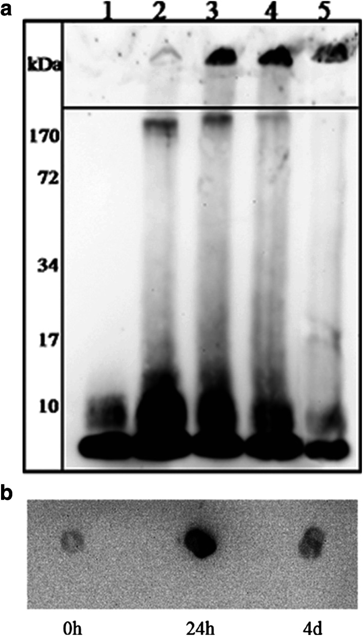
Fig. 1.
Visualization of Aβ(1–40) species during aggregation. a Western blot analysis of Aβ(1–40) samples, separated on a 12% bis–Tris SDS-PAGE and probed with 6E10 monoclonal antibody. Aβ(1–40) was incubated in TBS, pH 7.4 for 0 h (lane 1), 24 h (lane 2), 48 h (lane 3), 72 h (lane 4) and 16 days (lane 5). b Reactivity of Aβ(1–40) aggregates to A11 antibody. Dot blot analysis: Aβ(1–40) after 0 h, 24 h and 4 days in TBS, pH 7.4

