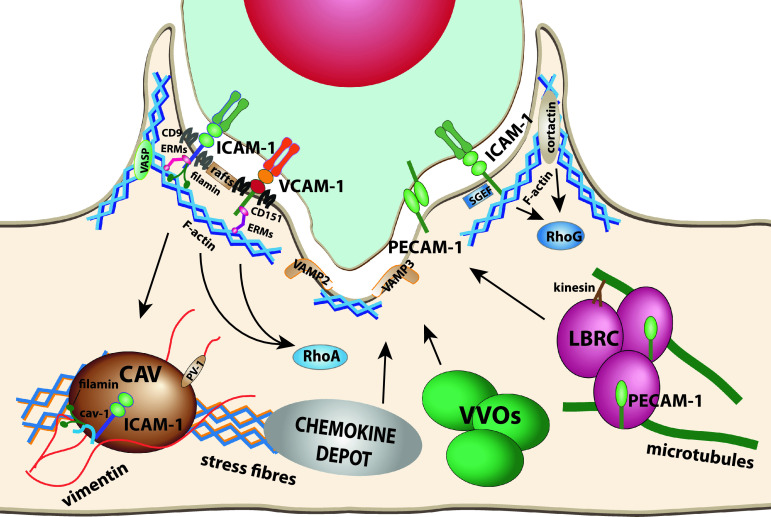
Fig. 1.
Proteins involved in endothelial apical membrane reorganization upon leukocyte interaction. ICAM-1, VCAM-1, and PECAM-1 are the three best characterized receptors that modulate the endothelial cell surfaces in contact with leukocytes. ICAM-1 and VCAM-1 are embedded into tetraspanin webs. Both receptors and E-selectin can be transiently recruited to membrane rafts upon engagement. ICAM-1 and VCAM-1 are connected to subcortical actin through different protein connectors such as ERM proteins or filamins. These receptors form signaling platforms in apical microvillar reorganization called docking structures. ICAM-1 and VCAM-1 signal to different Rho GTPases and cortactin to tune actin dynamics in the surrounding of endothelial cell-leukocyte contact areas and facilitate diapedesis. When apical ICAM-1 is engaged in caveola-rich cellular areas, can be internalized into basolateral caveolae. It has been proposed that PECAM-1 is translocated from the LBR compartment to endothelial contact areas. On the ventral side of the leukocyte, endothelial SNARE machinery participates in membrane remodeling in response to leukocytes emitting pseudopods/podosomes that scan for gateways to traverse the endothelial monolayer. VVOs participate in the formation of endothelial invaginations surrounding podosomes. Chemokine depots are intracellular compartments, still uncharacterized, which translocate chemokines to endothelial surface upon leukocyte adhesion. Actin stress fibers determine the localization of caveolae and chemokine depots. The vimentin network is associated with caveolae through PV-1. Microtubules and kinesins regulate LBRC dynamics

