Fig. 2.
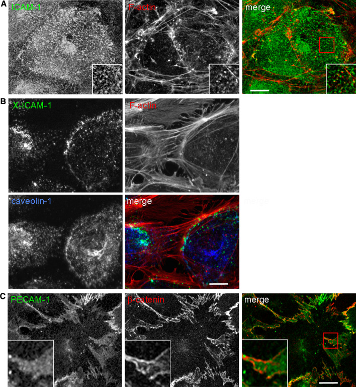
Endothelial membrane domains involved in adhesion receptor dynamics. a Microvilli. Confocal images of HUVECs stained for ICAM-1 and F-actin (phalloidin) showing the accumulation of this receptor in apical microvilli, involved in the formation of docking structures in response to leukocyte adhesion b Caveolae. Upon antibody-mediated ICAM-1 clustering (X-ICAM-1), actin stress fibers are increased, ICAM-1 aligns with actin filaments and partially colocalizes with caveolin-1. Leukocytes transmigrate transcellularly through areas rich in caveolin-1. c Lateral border recycling (LBR) compartment. PECAM-1 is diffusely localized at cell borders, labeled with anti-β-catenin antibody, in an internal compartment that is translocated to the cell surface upon leukocyte interaction. The LBR compartment is required for both paracellular and transcellular diapedesis. Bar 10 μm

