Fig. 2.
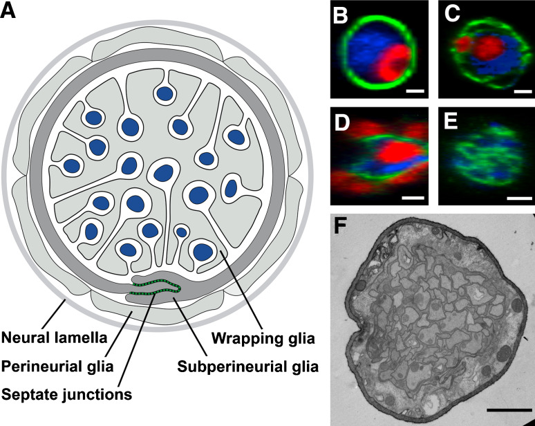
Drosophila peripheral nerve. a Schematic drawing of a cross-section of a third instar larval peripheral nerve. Perineurial glial cells are covered by an extracellular matrix called neural lamella. b–e Orthogonal cross-sections of segmental nerves. GFP-expression is shown in green, glial nuclei express Repo (red), axonal membranes are labeled by HRP staining (blue). b The neural lamella is marked by the GFP-gene trap insertion into the viking gene, which encodes Drosophila CollagenIV. c The perineurial glial cells are labeled in c527Gal4; UASCD8GFP flies. d The septate junction forming subperineurial glial cells express the moodyGal4 driver (genotype: moodyGal4; UASCD8GFP). e Wrapping glial cells are marked by expression of the nervana2Gal4 driver (genotype: nrv2Gal4; UASS65TGFP). f Electron micrograph of a cross-section through a larval peripheral nerve at the third instar stage. Scale bars 2 μm

