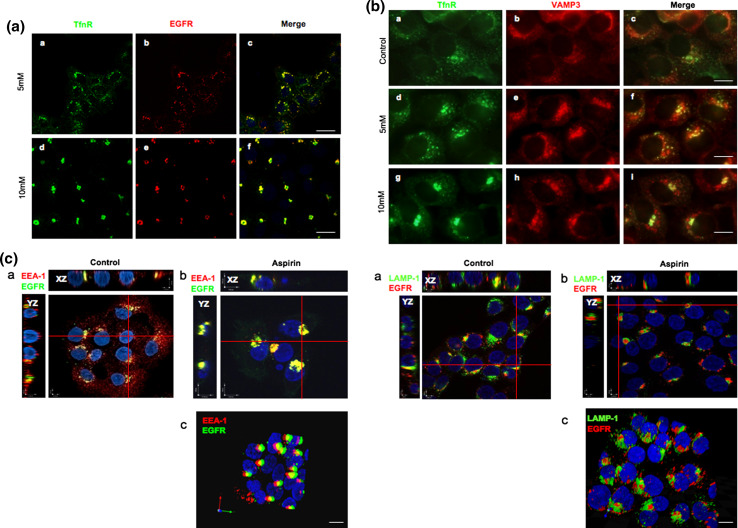
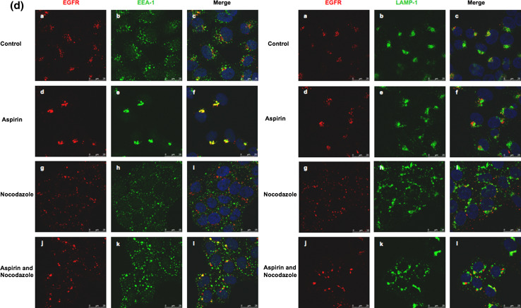
Fig. 3.
AMC is part of the early endosomes. a Cells were serum starved for 12 h prior incubation with Alexa Fluor 555-conjugated EGF and anti-TfnR antibodies for 1 h on ice. Thereafter, cells were treated with varying concentrations of aspirin for 2 h. Endocytosed-TfnR was stained with secondary antibody. Image of confocal microscope, scale bars 50 μm. b Cells were treated as in a , followed by staining with polyclonal anti-VAMP-3 primary antibody and secondary antibodies. Image of fluorescence microscope, 100×. c Cells were serum starved for 12 h prior to incubation with Alexa Fluor 555-conjugated EGF for 1 h on ice. Thereafter, cells were treated with 10 mM aspirin for 2 h, stained with anti-EEA-1 (left) or anti-LAMP-1 (right) antibodies for 1 h, followed by secondary antibodies. Image of X–Z/Y–Z projection using confocal microscope (a, b). Image of stereo three-dimensional, confocal microscope (c). d Cells were serum starved for 12 h prior to incubation with Alexa Fluor 555-conjugated EGF for 1 h on ice. Thereafter, cells were treated with and without 10 mM aspirin for 1 h, followed by treatment with and without 10 μg/ml nocodazole in the presence or absence of aspirin for 1 h. Image of confocal microscope, scale bars 25 μm. The colocalizations of internalized EGF with EEA-1 and LAMP-1 in various situations are indicated by Pearson’s correlation (r). r average of values from five frames per coverslip in a representative of two independent experiments. In EEA-1 staining, control = 0.22; aspirin = 0.42; nocodazole = 0.17; aspirin and nocodazole = 0.33. In LAMP-1 staining, control = 0.31; aspirin = 0.26; nocodazole = 0.30; aspirin and nocodazole = 0.29


