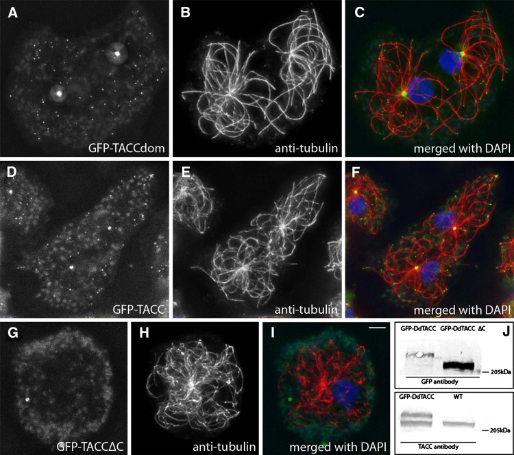
Fig. 5.
The TACC domain is sufficient for localization at centrosomes and microtubule tips. GFP-TACCdom (a–c) and the full-length GFP-TACC (d–f) localize to the centrosomal corona, microtubule plus-end tips and to certain spots alongside of microtubules. By contrast, a C-terminally truncated fusionprotrein GFP-TACC∆C (g–i) lacking the TACC domain does not show any specific localization. GFP labeling (a, d, g) is shown in green, tubulin (b, e, h) in red and DNA in blue. Except for GFP-TACC∆C (i), merged images (c, f) clearly show the GFP-TACC and GFP-TACCdom spots at the microtubule tips. The Western blot of Dictyostelium extracts (j) shows labeling of GFP-TACC and GFP-TACC∆C with anti-GFP (upper panel) and of GFP-TACC and endogenous TACC with anti-TACC (lower panel). Bar 2 μm

