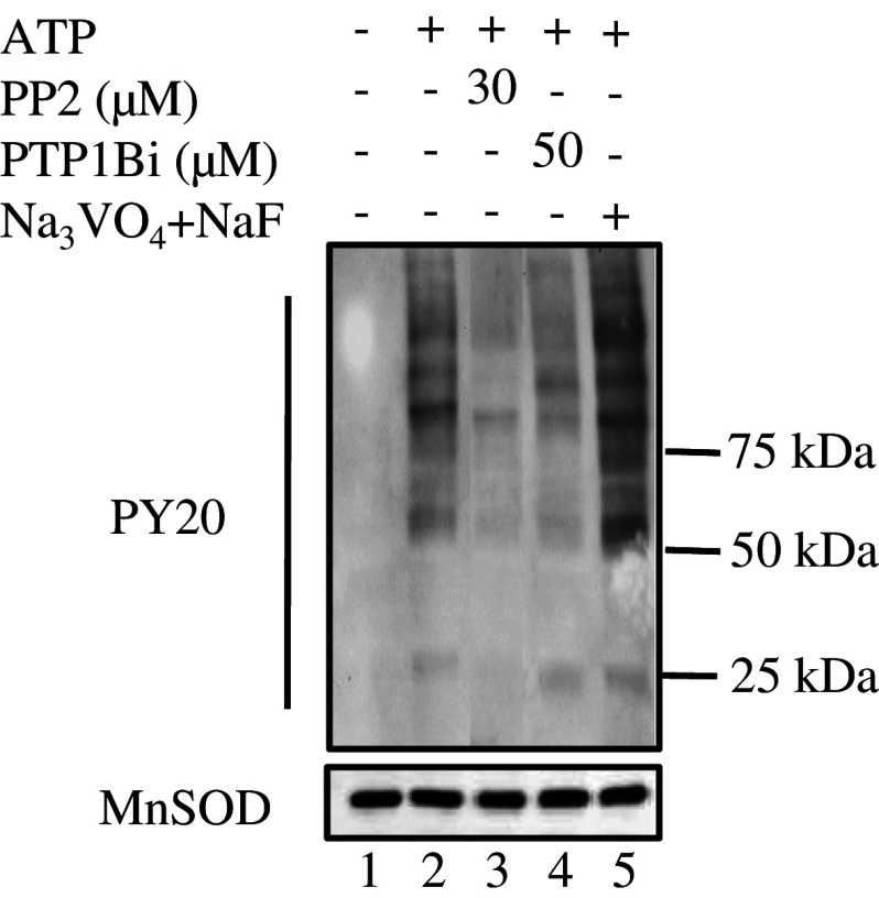
Fig. 1.
Changes in protein tyrosine phosphorylation of rat brain mitochondria following exposure to ATP, PP2, PTP1Bi, or orthovanadate + NaF. Mitochondria (50 μg) were untreated (lane 1) or treated at 30°C for 10 min with 1 mM ATP alone (lane 2) or after pre-incubation at 30°C for 10 min with 30 μM PP2 (lane 3), 50 μM PTP1Bi (lane 4), or 2 mM orthovanadate plus 10 mM NaF (lane 5). The reaction was stopped by addition of 5× Laemmli buffer. Tyrosine-phosphorylated proteins on immunoblots were detected with an antibody to phosphotyrosine. The membrane was stripped and reprobed with an antibody to MnSOD as a loading control (below). Data are representative of at least three experiments

