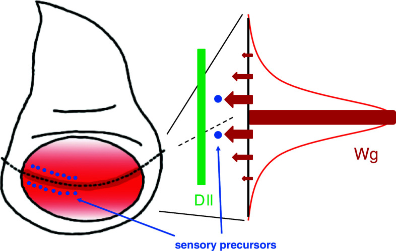
Fig. 2.
Wg morphogen in the Drosophila wing disc. (Left) Wg is expressed in cells (dark red) at the dorsoventral boundary (a dotted line). Secreted Wg is distributed in a concentration gradient manner (red). Sensory organ precursor cells (blue dots) are induced near the Wg-expressing region. (Right) Different levels of signal strength (arrows) are evoked depending on extracellular Wg concentration (red). A strong signal induces sensory precursor cells (blue dots) in the proximal region to the Wg-expressing cells. Dll expression (green) is induced over a broad area because it requires a lower threshold of Wg concentration

