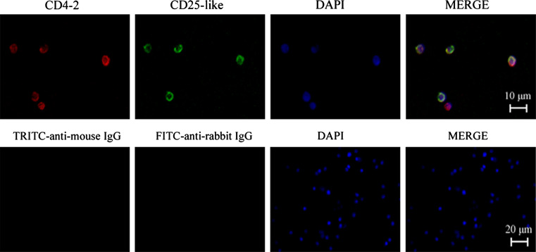
Fig. 3.
Immunofluorescence stains of CD4-2+CD25-like+ leucocytes. CD4-2 molecules were detected with TRITC-conjugated anti-mouse IgG that revealed red fluorescence, while CD25-like molecules were detected with FITC-conjugated anti-rabbit IgG that exhibited green fluorescence. DAPI stain shows the location of the nuclei. The negative controls are shown in the second row, and consisted of negative mouse and rabbit IgGs in place of primary anti-CD4-2 and anti-CD25-like Abs

