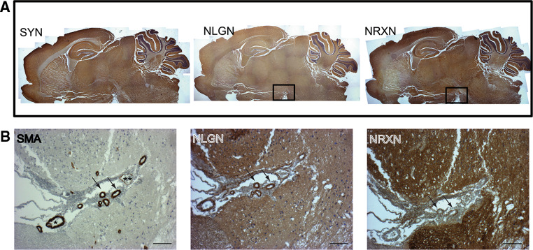
Fig. 2.
Neurexin and Neuroligin expression in blood vessels of the adult mouse brain. a The three panels were produced by merging several low magnification images taken from a sagittal section of a mouse brain stained with antibodies to Synaptophysin, Neurexin and Neuroligin, which globally produce a similar expression pattern. Enriched areas for Neurexin and Neuroligin expression are located in the cortex, hippocampus, hypothalamus and cerebellum. b Magnifications of an area proximal to the hypothalamus (boxed area in a), which presents many large vessels, including arteries and veins. An artery (*) and a vein (**) are highlighted. Arrows indicate blood vessels in which Neurexin expression is limited to the outer layer of the vessel wall. Scale bar 100 μm. This figure was partially modified from ref. [80]

