Fig. 5.

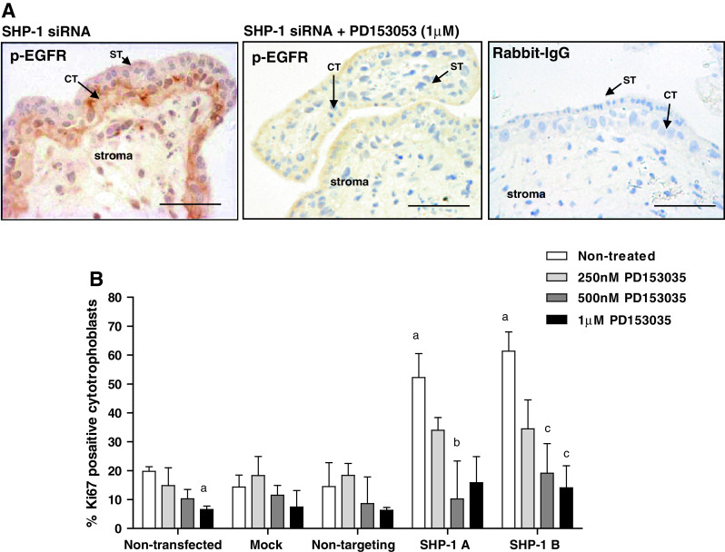
Enhanced proliferation following SHP-1 knockdown can be attributed to enhanced EGFR activation. First-trimester placental explants were transfected with non-targeting siRNA (500 nM) or SHP-1 siRNA (500 nM) for 48 h in serum-free conditions. Explants were exposed to the EGFR-specific inhibitor PD153035 (250 nM to 1 μM) for a further 24 h then EGFR activation (a) and cytotrophoblast proliferation (b) was assessed by immunohistochemistry. Three random areas from each placenta were counted, and the number of Ki67-positive cells is expressed as a percentage of the total number of cytotrophoblast (median and IQR) of at least five independent experiments (b). Kruskal–Wallis test of variance followed by Dunn’s multiple comparison post hoc test was used to assess significant (p < 0.05) differences between the groups. a Significantly different from non-treated, non-transfected; b, significantly different from non-treated, SHP-1A siRNA transfected; c significantly different from non-treated, siRNA B siRNA transfected. ST syncytiotrophoblast, CT cytotrophoblast. Scale bars 50 μm
