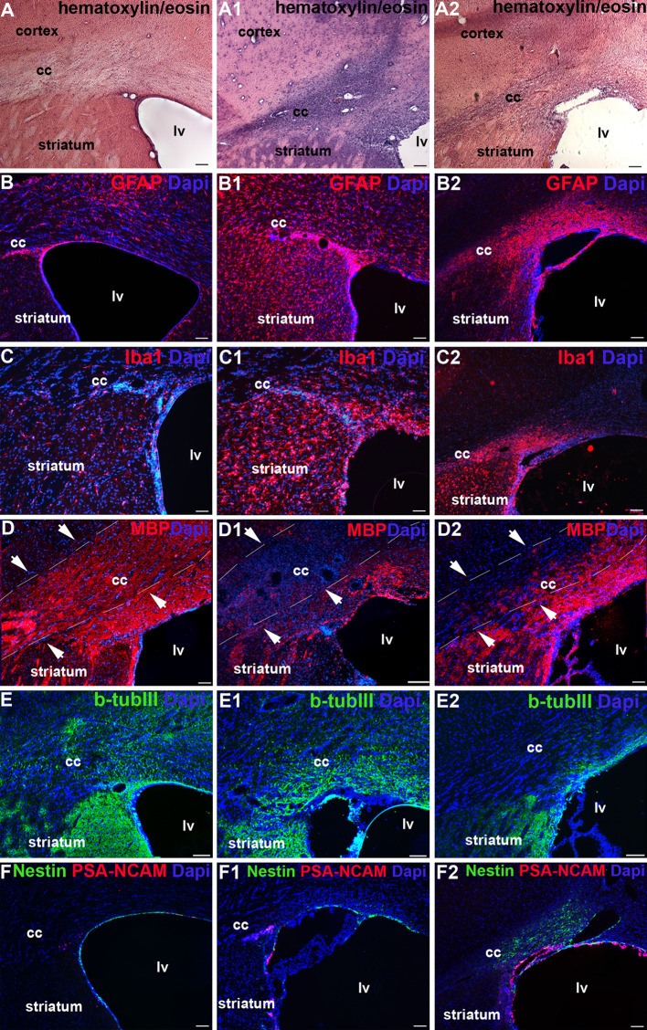
Fig. 1.
Inflammatory reaction and tissue damage after LPC lesion: Analysis of cc in control (not lesioned) rats (a–f), and animals lesioned at 5 (a1–f1) and 20 (a2–f2) days after LPC injection. Immunohistochemistry: a–a2 hematoxylin and eosin; b–b2 GFAP; c–c2 Iba1; d–d2 MBP; e–e2 β-TubIII; f–f1 nestin/PSA-NCAM. Total nuclei are shown by DAPI staining (blue). Scale bars, in a–f2 50 μm. lv Lateral ventricle, cc corpus callosum

