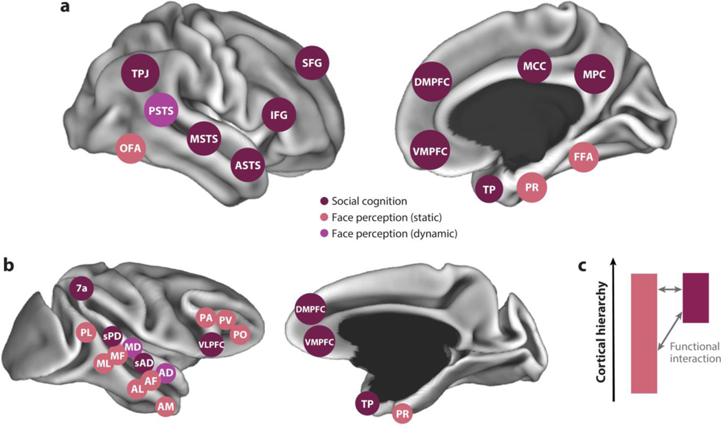
Figure 1.
Areas of functional specialization for social cognition in (a) humans and (b) macaques shown from a lateral view (left) and medial view (right). Areas specialized for static and dynamic face perception (salmon and purple, respectively) and high-level social cognition (burgundy), including theory of mind, are indicated. Areas are shown schematically on computer-rendered cortical surfaces. (c) Systems for face perception and social cognition occupy distinct regions of the primate anatomical connectome, but they interact functionally. Abbreviations: AD, anterior dorsal; AF, anterior fundus; AL, anterior lateral; AM, anterior medial; ASTS, anterior STS; DMPFC, dorsomedial prefrontal cortex; FFA, fusiform face area; IFG, inferior frontal gyrus; MCC, middle cingulate cortex; MD, middle dorsal; MF, middle fundus; ML, middle lateral; MPC, medial parietal cortex; MSTS, middle STS; OFA, occipital face area; PA, prefrontal arcuate; PL, posterior lateral; PO, prefrontal orbital; PR, perirhinal cortex; PSTS, posterior superior temporal sulcus; PV, prefrontal ventral; sAD, social anterior dorsal; SFG, superior frontal gyrus; sPD, social posterior dorsal; STS, superior temporal sulcus; TP, temporal pole; TPJ, temporo-parietal junction; VLPFC, ventrolateral prefrontal cortex; VMPFC, ventromedial prefrontal cortex.

