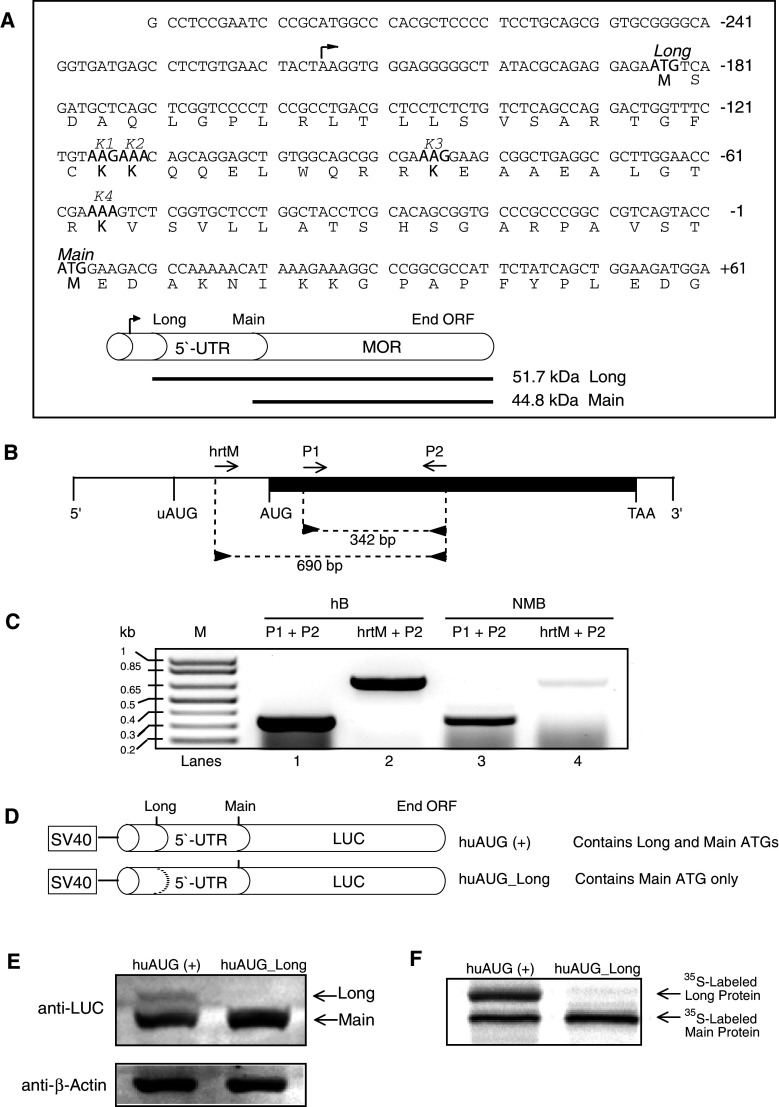
Fig. 1.
Schematic representation of the human μ opioid receptor (OPRM1) and putative 5′-UTR sequences. a Sequence of the OPRM1 5′-UTR used in this study. The in-frame uAUG (Long) and main AUG (Main) codons are indicated, as are the peptide sequences for the lysine (K1, K2, K3, and K4) codons. The arrowhead above the sequence indicates the transcription initiation site. The upstream and main AUGs encode the Long and Main forms of OPRM1, respectively. b Reverse transcription (RT)-PCR was carried out using oligonucleotide primers (“Materials and methods”). The diagram shows the relative position of sense (hrtM and P1; forward arrows) and antisense (P2; reverse arrows) primers along the OPRM1 mRNA. The darkened area represents the exon region (1,403 bp). Vertical lines represent the relative positions of the common 5′ end, initiation codon for the Long form of OPRM1 (uAUG), initiation codon for the Main form of OPRM1 (AUG), termination codon for both OPRM1s (TAA), and the 3′ end. c RT-PCR analysis of the OPRM1. Total RNA prepared from human brain (hB, lanes 1 and 2; Ambion) and NMB cells were subjected to RT-PCR with the oligonucleotide primer pairs indicated above the lanes followed by agarose gel electrophoresis. Sizes of amplification products obtained with the indicated primer pairs are shown. M denotes DNA markers, the sizes of which are shown on the left. d Luciferase fusion constructs containing portions of OPRM1 extension sequences. The huAUG (+) construct retains both the 5′ in-frame and main AUGs; the huAUG_Long construct retains only the main AUG. The dotted line represents ATGs converted to ACGs by point mutations. e NMB cells were transfected transiently with the constructs shown in d, and protein levels were analyzed by Western blotting for luciferase and β-actin. f Representative autoradiogram of proteins translated in vitro in the presence of [35S]-methionine using a coupled transcription/translation system

