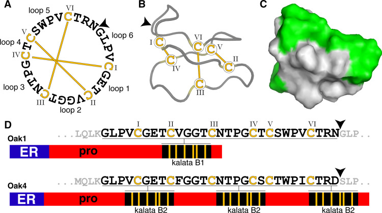
Fig. 1.
Schematic overview of the prototypic cyclotide kalata B1 and two precursor proteins, Oak1 and Oak4. (Oak refers to O ldenlandia a ffinis kalata). a Sequence of kalata B1 showing disulfide connectivities, loops between conserved Cys residues and the ligation point (arrow) involved in peptide cyclisation. b A ribbon model of kalata B1 highlighting the CCK motif of three disulfide bonds and cyclic backbone. c A surface-rendered model of kalata B1 showing the hydrophobic patch (green) which associates with membranes. d Precursor proteins for kalata B1 (Oak1) and kalata B2 (Oak4). Oak4 is shown to highlight the fact that some cyclotide genes encode multiple copies of cyclotides (as is the case for kalata B2) [16]. The cyclotide C-terminal residue (Asn or Asp), thought to be targeted by AEP, is marked with an arrow

