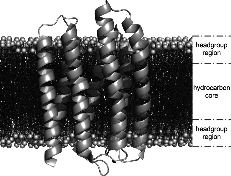
Fig. 1.
Seven-helix phototaxis receptor sensory rhodopsin II in a phospholipid bilayer. A high-resolution protein structure (PDB 1jgj) obtained by X-ray crystallography [182] was superimposed onto a bilayer manually assembled from 3,648 1-palmitoyl-2-oleoyl-sn-glycero-phosphocholine (POPC, Molfile from Avanti Polar Lipids, Alabaster, AL) monomers. Each of the two headgroup regions is about 15 Å thick, whereas the hydrocarbon core is about 30 Å in thickness

