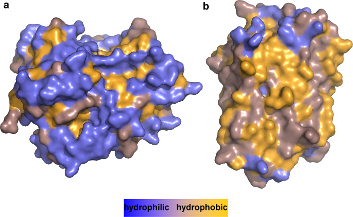
Fig. 4.
Differences in the hydrophobicity of surface areas between globular water-soluble and α-helical membrane proteins of similar size. Colour code is blue for hydrophilic surfaces, orange for hydrophobic ones and dark salmon for residues that are in between. a Human carbonyl reductase 1 (PDB 1wma, 276 residues) presents mainly hydrophilic residues to its aqueous environment and shields hydrophobic ones in its core. b Bacteriorhodopsin from Halobacterium salinarum (PDB 1c3w, 231 residues) exposes mostly hydrophobic residues to its lipid bilayer environment, whereas hydrophilic residues are found in the rather small regions that are in contact with lipid headgroups and water. Note that the interiors of membrane proteins are as hydrophobic as those of water-soluble proteins (not shown, see [20, 22])

