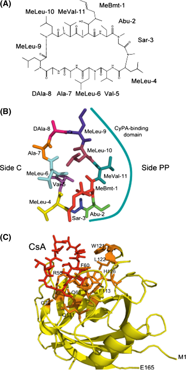
Fig. 1.
Cyclosporine-A. a The chemical structure of CsA with the following abbreviations: Abu L-α-aminobutiric acid, MeBmt (4R)-4[(E)-2-butenyl]-4,N-dimethyl-L-threonine, MeLeu N-methylleucine, MeVal N-methylvaline, Sar sarcosine. b CsA structure extracted from its complex with hCyPA (1CWA.pdb); the side interacting with CyPA (side PP) is marked with the solid line and side C with the residues interacting with calcineurin. c Full structure of the hCyPA/CsA complex (1CWA.pdb); CsA is marked as red sticks, the AA residues forming the PPIase cavity are shown as orange sticks while the remaining backbone of the cyclophilin is in yellow. M1 N-terminal methionine, E165 C-terminal glutamic acid

