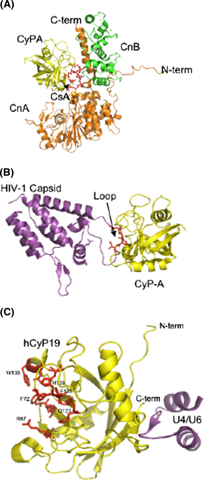
Fig. 6.
Three X-ray structures of the binary and tertiary complexes; a the (hCyPA/CsA + CnA/CnB) complex [13]; CyPA (yellow) binds to CsA (red, indicated with a black arrow) and interacts with CnA (orange ribbons) and CnB (green ribbon) complex. b X-ray structure of HIV-1 capsid protein (violet) bound to the PPIase cavity of hCyPA (yellow ribbon); the loop in red comes from the capsid protein (1M9C.pdb, [30]). c X-ray structure of a short peptide from the U4/U6 snRNP-60K (violet) protein interacting with the hCyP19 (yellow ribbon) with some of the AAs forming its PPIase cavity (red sticks) (1MZW.pdb, [18])

