
Fig. 1.
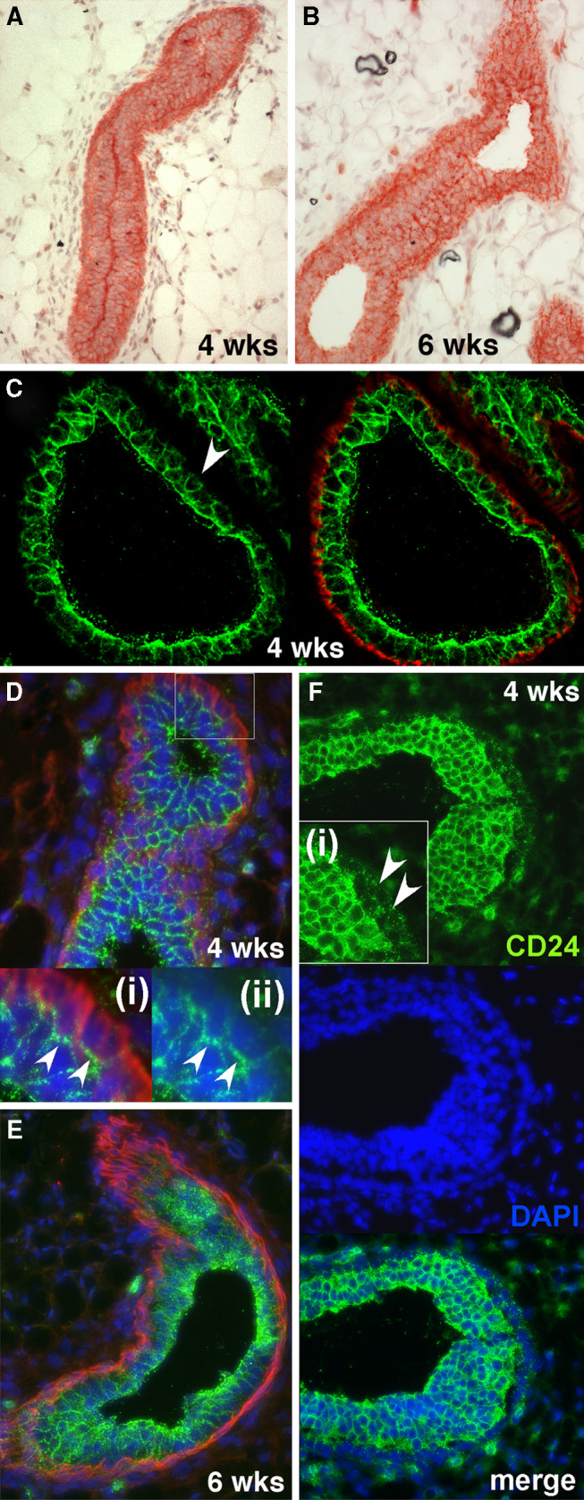
Expression of CD24 in the murine mammary gland during puberty. a, b CD24 expression detected by Fuchsin chromogen staining in mammary epithelial ducts from mice 4 weeks old (a) and 6 weeks old (b). c–e Expression of CD24 (green fluorescence) and smooth muscle actin (red fluorescence) in mammary glands from 4-week-old mice (c, d) and 6-week-old mice (e). DAPI staining of nuclei (blue fluorescence) was omitted from c for the sake of clarity. The arrowhead in c indicates an example of a luminal cell with only weak CD24 staining on its basally-facing surface. The insets (i) and (ii) in d show larger magnifications of the boxed area in the main picture. In inset (ii) the smooth muscle actin staining has been omitted to allow better visualization of the strong CD24 staining at the interface between luminal and basal cells (indicated by arrowheads) and the weak CD24 staining elsewhere on the basal cells. f CD24 staining (green fluorescence) and DAPI staining of nuclei (blue fluorescence) of a TEB from mice of 4 weeks of age. The arrowheads in inset (i) indicate weak CD24 staining in the cap cells of the TEB