
Fig. 1.
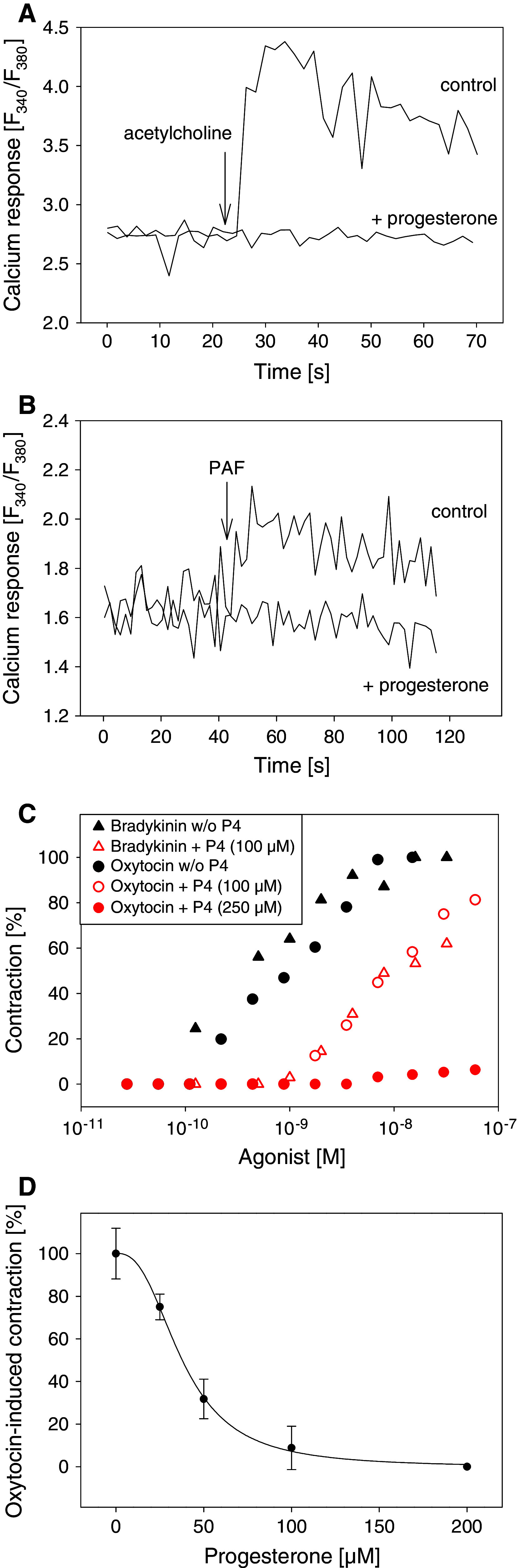
Progesterone inhibits cellular calcium responses and agonist-induced contractions of the uterus. a Fura-2-loaded HEK293 cells were treated with 160 μM progesterone or ethanol (control) for 20 min at 37°C. Calcium measurements were started and 100 μM acetylcholine was added (arrow). b Fura-2-loaded MFE-280 cells were treated with 160 μM progesterone or ethanol (control) for 20 min at 37°C. Calcium measurements were started and 6 μM platelet activating factor (PAF) was added (arrow). a, b Representative calcium traces. c Rat uteri were treated with progesterone (P4) or vehicle (30°C, 3 min). The contraction height as response to the indicated concentrations of the corresponding ligands was measured. The 100% level is equal to the maximum contraction obtained with the indicated ligands in the absence of progesterone. d Rat uteri were treated with different concentrations of progesterone (30°C, 3 min) and then oxytocin was added. The contraction height as response to 0.5 nM oxytocin was measured. Curve fitting was performed using a four-parameter logistic function (IC50 = 37 μM). Standard deviations were calculated from oxytocin-induced contractions from 2–4 different strips