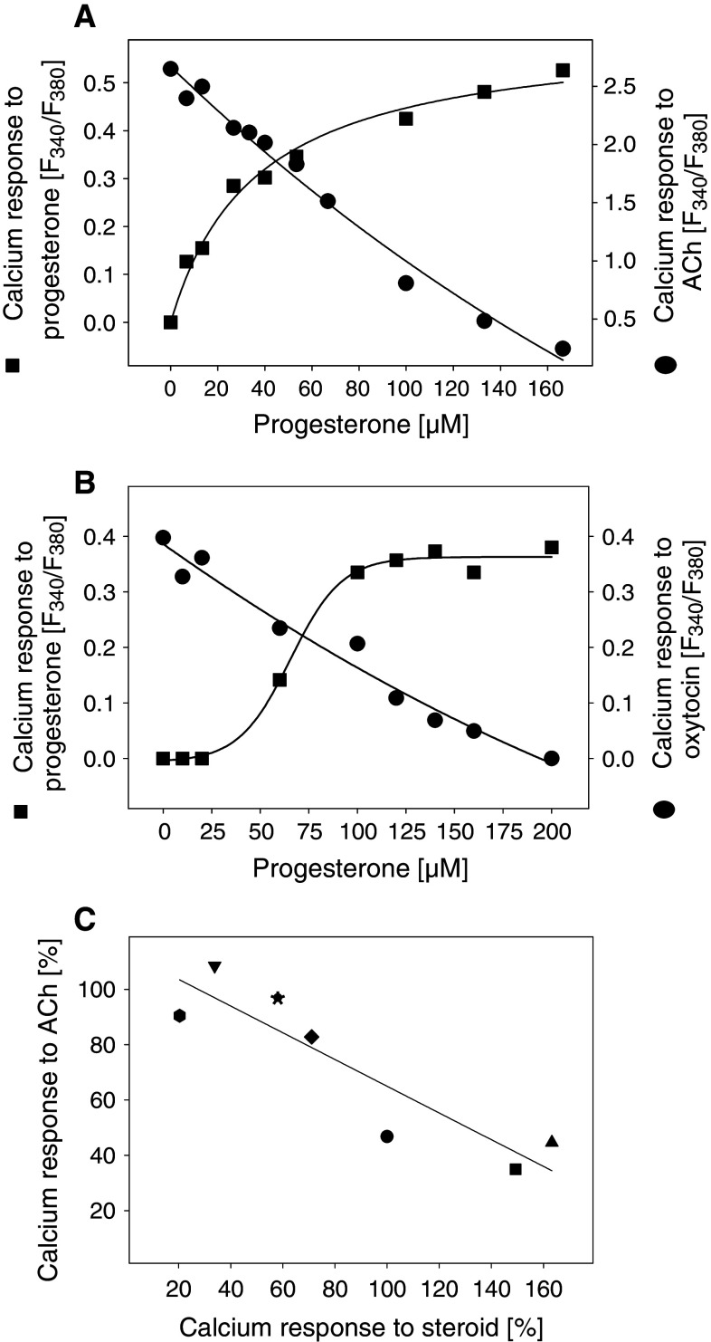
Fig. 6.
Correlation of cellular calcium increase evoked by steroids with the inhibition of calcium response to acetylcholine (ACh) or oxytocin. Calcium responses of fura-2-loaded HEK293 cells (a) or myometrial PHM1-31 cells (b) to progesterone were measured (squares). Three min after the addition of progesterone, calcium responses to 100 μM ACh (a) or 15 nM oxytocin (b) were analyzed (circles). c Abscissa: Calcium responses of fura-2-loaded HEK293 cells to different steroids (100 μM each). The calcium response to progesterone was set to 100%. Ordinate: After 3 min incubation with 100 μM steroid at 37°C, the calcium response to 100 μM ACh was measured. The calcium response of ethanol-treated control cells was set to 100%. Filled circle Progesterone, filled square 5β-dihydroprogesterone, filled uptriangle pregnenolone, filled downtriangle hydrocortisone, filled diamond DHEA, filled hexagon 11α-hydroxyprogesterone, filled asterisk 11β-hydroxyprogesterone

