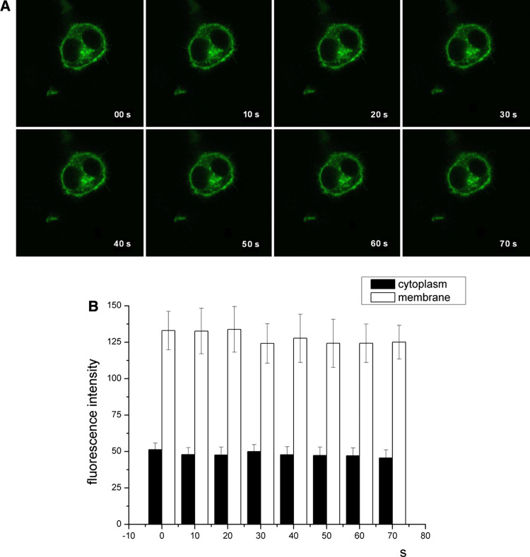
Fig. 2.
a Confocal images of HEK 293 cells transiently transfected with rGAT1-GFP. The fluorescent signal at the plasma membrane is relatively stable over time, before and after imipramine treatment: imipramine 100 μM was added between image at t = 0 and image at t = 10. b Mean fluorescence intensity measured at plasma membrane (white columns) and cytoplasm (black columns). Error bars ± SE, n = 4

