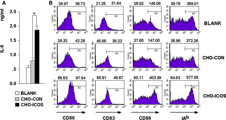
Fig. 4.
Membrane-anchored ICOS induces expression of IL-6 by DCs. DCs were left untreated, or cocultured with CHO-ICOS or CHO-CON which were pre-treated by mitomycin for 24 h. a The supernatant were collected and detected for IL-6 with ELISA kit (*P < 0.05). b Cells were then collected and washed, treated with different fluorescence-labeling antibody and detected by FCM; CD11c+ cells were gated and the data were analyzed by Cellquest Software. Left-hand numbers above each histogram represent the percent of positive-staining cells while the right-hand numbers represent mean fluorescent intensity (MFI) of all the cells gated (data are one representative of three)

