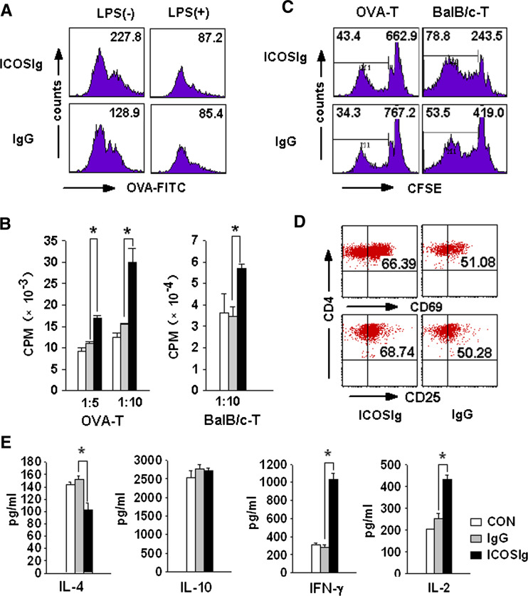
Fig. 7.
ICOSIg induces immunogenic DCs in vitro. a Immature DCs were treated with ICOSIg or IgG1 in the absence (left) or presence (right) of 100 ng/ml LPS for 24 h, then washed thoroughly and cocultured with 100 μg/ml FITC labeling OVA protein and detected by FCM. Numbers shown represent MFI of all the cells gated. b–e Immature DCs were pretreated by ICOSIg. b Cocultured with OVA-specific CD4+ T in the presence of OVA323–339 peptides (left) or with CD4+ T obtained from spleen of BALB/c mouse (right) for another 72 h and H3-thymine was added before the last 18 h. X-axis gives the ratio of DC: T(*P < 0.05). c Cocultured with CD4+ T prestained by CFSE agent for 5 days and analyzed by FCM. Left-hand numbers represent the percent of offspring T cells after more than three times of cell division, right-hand numbers represent the MFI of all the cells gated. d Cocultured with OVA-specific CD4+ T as in (b). T cells were then washed and evaluated the expression of CD25 and CD69. e Cocultured with OVA-specific CD4+ T as in (d). The supernatant of the coculture system were then collected and assayed for different cytokines with corresponding ELISA Kit (*P < 0.05)

