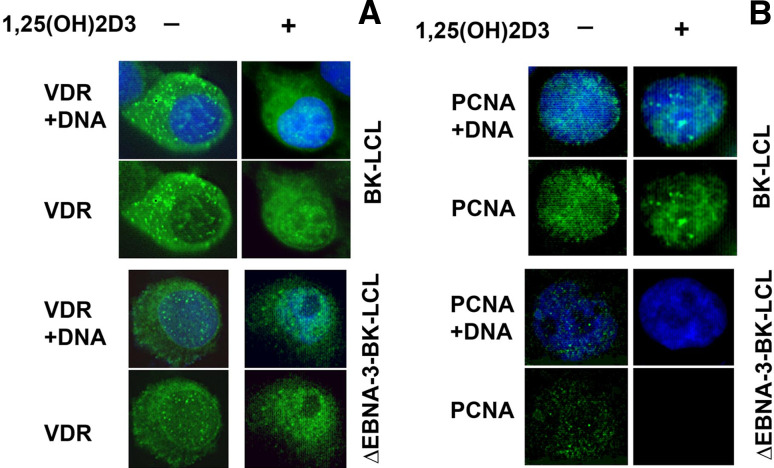
Fig. 6.
Immunostaining of BK-LCL and ∆EBNA3-BK-LCL cells, untreated and treated with 1,25(OH)2D3. a Staining with anti-VDR antibody. Notice that upon treatment with the ligand VDR translocates to the nucleus regardless of the presence of EBNA-3. b Staining for proliferating cell nuclear antigen, PCNA. Notice that upon the ligand treatment there is no PCNA signal in the nucleus of ∆EBNA3-BK-LCL cells, in contrast to EBNA-3 expressing LCL

