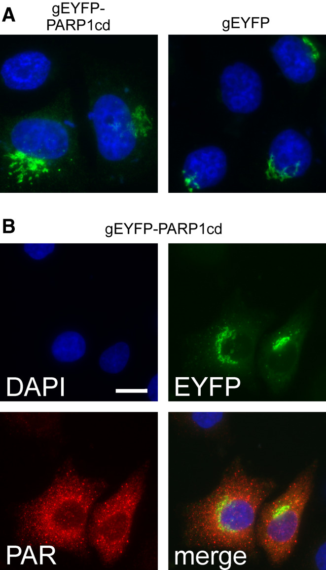
Fig. 5.
Golgi-targeted PARP1cd fusion proteins give rise to PAR-positive cytoplasmic vesicles. a The Golgi-targeted EYFP-PARP1cd fusion protein exhibits the same subcellular distribution as an established Golgi-EYFP construct carrying the same targeting sequence. The proteins were detected by their intrinsic fluorescence, nuclei were stained with DAPI. b HeLa S3 cells expressing a Golgi-targeted PARP1cd fusion construct displayed consistent PAR immunoreactivity as revealed by immunocytochemistry. While the protein was detected in Golgi structures by its intrinsic EYFP fluorescence, the PAR signal most often did not co-localize with the EYFP signal, but was detected in cytoplasmic vesicles. Note that in part of the transfected cells both the protein and the PAR co-localized within the Golgi complex (cf. Fig. 3b). Bar 10 μm

