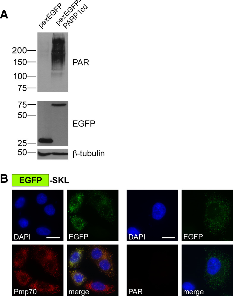
Fig. 6.
Detection of peroxisomal protein localization. a PAR immunoblot analysis of lysates from HeLa S3 cells transiently expressing EGFP or EGFP-PARP1cd targeted to peroxisomes as indicated. PAR accumulation was detected only in lysates from cells expressing the pexEGFP-PARP1cd protein. Each lane was loaded with 70 μg of total protein. The overexpressed proteins were detected using an antibody recognizing the EGFP portion, β-tubulin served as loading control. b HeLa S3 cells transiently expressing EGFP targeted to the peroxisomes by a C-terminal SKL sequence were subjected to immunocytochemistry to detect the endogenous marker Pmp70 (left) or PAR (right). As monitored by its intrinsic EGFP fluorescence the protein co-localized with peroxisomal structures (Pmp70). However, the cells were negative for PAR immunoreactivity. Bar 10 μm

