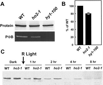
Figure 6.
Detection of apo-phyA in ho2-1 seedlings. A, Detection of the phyA protein and bound PΦB chromophore in wild-type ecotype WS (WT), hy1-100, and ho2-1 seedlings. Crude extracts were prepared from 4-d-old etiolated seedlings and subjected to immunoblot analysis with the anti-phyA monoclonal antibody O73D (top) or assayed for bound PΦB by zinc-induced fluorescence (lower). B, Quantitation of the levels of bound PΦB per phyA protein. Signals obtained from A were quantitated by densitometric scans of the gels. Values (±sd) were expressed relative to those from WT. C, R-light induced degradation of phyA in etiolated seedlings of WT and ho2-1. Etiolated seedlings were irradiated continuously with R. At various times, crude extracts were prepared and subjected to immunoblot analysis with the anti-phyA monoclonal antibody O73D; an equal amount of seedlings (grams fresh weight) was analyzed for each sample.

