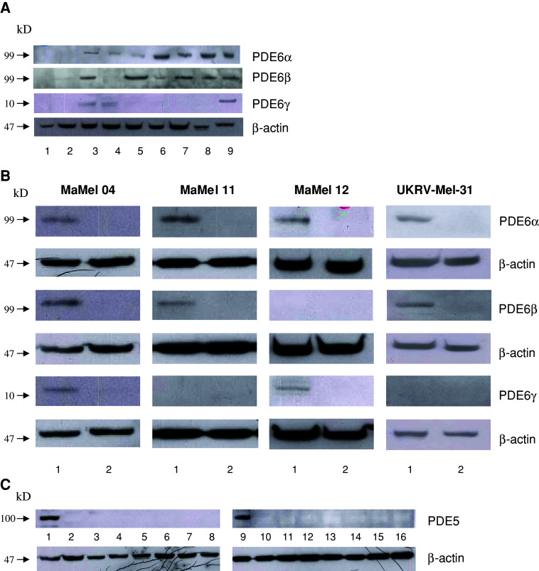
Fig. 1.
Protein expression of PDE6 and PDE5. a Expression of PDE6 subunits in NEM (1), HaCaT (2) and melanoma cell lines Ma-Mel-04 (3), Ma-Mel-12 (4), UKRV-Mel-31 (5), Ma-Mel-11 (6), Ma-Mel-21 (7), and MeWo (8), as well as Y79 retinoblastoma cell line as a positive control (9); b expression of PDE6 subunits in untransfected controls (1) or in cell lines transfected with shDNA plasmids (2) as indicated on the right side; c expression of PDE5 in heart as a positive control (1, 9), NEM (2), HaCaT (3), and melanoma cell lines Ma-Mel-04 (4), Ma-Mel-12 (5), UKRV-Mel-31 (6), Ma-Mel-11 (7), Ma-Mel-21 (8), MeWo (10), UKRV-Mel-06 (11), UKRV-Mel-14a (12), UKRV-Mel-21 (13), SK-Mel23 (14), Ma-Mel-05 (15), and Ma-Mel-21 (16)

