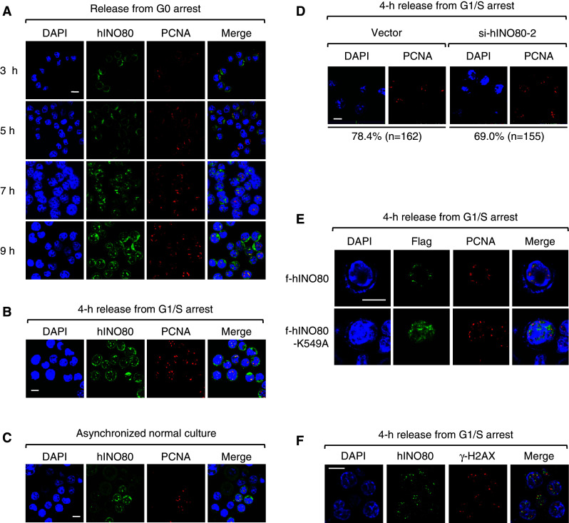
Fig. 5.

hINO80 is co-localized with PCNA at replication forks during the S-phase. a Sub-confluent HeLa cells were arrested at G0 by serum starvation, and after release for various times, the cells were fixed and dually stained with the antibodies for hINO80 and PCNA. Representative confocal images are shown. Quantitation of colocalization only within the nucleus using the LSM510 META-3.2 software indicates that hINO80 and PCNA overlaps by 90% at the 9-h time point. Note that the cells gradually become larger in size as the cell cycle moves forward into the S-phase. b HeLa cells were released from G1/S arrest for 4 h and fixed for staining with the antibodies for hINO80 and PCNA before confocal images were captured. Quantitation of colocalization between hINO80 and PCNA done as in (a) indicates that they overlap by 90%. c Asynchronized normal HeLa cells were fixed and dually stained with the antibodies for hINO80 and PCNA. Confocal images of the population containing S-phase cells were captured. d Indicated cells were released from G1/S arrest for 4 h and fixed for staining with the PCNA antibodies. The percentages of PCNA focus forming cells were determined by counting the indicated number of cells. e 293T cells were transfected with the expression vectors for f-hINO80 or f-hINO80-K549A and arrested at G1/S. After release for 4 h, cells were fixed for staining with the antibodies for PCNA and Flag. f HeLa cells were arrested at G1/S and released for 4 h before cells were fixed and dually stained with the antibodies for hINO80 and γ-H2AX. Representative confocal images of each experiment are shown
