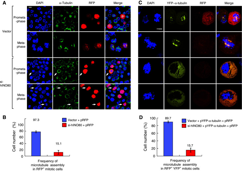
Fig. 7.

hINO80 is required for microtubule assembly during mitosis. a 293T cells were cotransfected with the RFP vectors plus either empty or si-hINO80 vectors at the molar ratios of 1:10. After release from G2/M arrest for 30 min (prometaphase) or 60 min (metaphase), cells were fixed for staining with the α-tubulin antibodies and DAPI. Representative confocal images are shown. The arrows indicate the RFP-positive cells defective in microtubule assembly during mitosis. b Data from the experiments in (a) were depicted as a graph. The frequency of microtubule assembly in RFP-positive mitotic cells was presented as mean ± SD from two independent experiments. Approximately 100 RFP-positive cells were counted for each sample. c 293T cells were cotransfected with the three different plasmids, the RFP and pYFP-α-tubulin vectors plus either empty or si-hINO80 vectors at the ratios of 1:1:10, and arrested at G2/M and released as in (a). Representative confocal images are shown. d Data from the experiments in (c) were depicted as a graph. The frequency of microtubule assembly in RFP/YFP double-positive mitotic cells was presented as mean ± SD from two independent experiments. Approximately 60 RFP/YFP double-positive mitotic cells were counted for each sample
