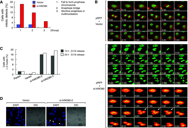
Fig. 8.

hINO80 deficiency causes abnormal chromosome segregation and multinucleation. a The GFP-H2B-expressing HeLa cells were transfected with the RFP vectors together with either empty or si-hINO80 vectors at the molar ratio of 1:10. Confocal live-cell images were captured every 5 min after release from G1/S arrest. Approximately 80 RFP-positive cells were evaluated for mitotic defects and the results were presented as a graph. b Representative confocal images are shown for the cells transfected with the RFP vector plus either empty (top) or si-hINO80 vectors (middle and bottom). The middle panel is a representative of the cells showing anaphase bridge (followed by chromosome fragmentation and cell death in this particular case), and the bottom is a representative of the cells showing abortive anaphase (indicated by arrows); accompanied by abnormal chromosome segregation into three in this particular case. The images at 10-min intervals were arranged so as to span the entire mitosis (the cells presumably starting to enter prophase were labeled by 0:00 time). c G1/S-arrested cells were released for 10 or 25 h, fixed and stained with DAPI. Percentages of the cells containing two or more nuclei were determined by counting 500–600 cells and depicted as a graph. d Representative confocal images used for the quantitation in (c) are shown. The cells containing two or more nuclei are indicated by arrows
