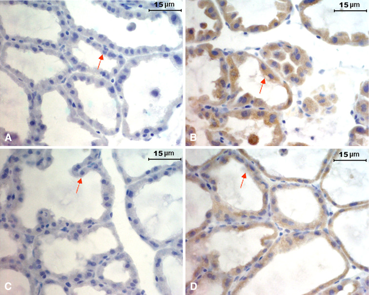
Fig. 1.
Expression of IgG in mammary gland epithelial cells. Immunohistochemical analysis using biotin-conjugated goat anti-mouse IgG and HRP-conjugated streptavidin showing positive staining in mammary gland epithelium of pregnant mice (day 17, b) and lactating mice (day 1, d). No staining in mammary gland epithelium of pregnant (a) and lactating mice (c) when using the goat IgG as isotype control. Scale bar 15 μm

