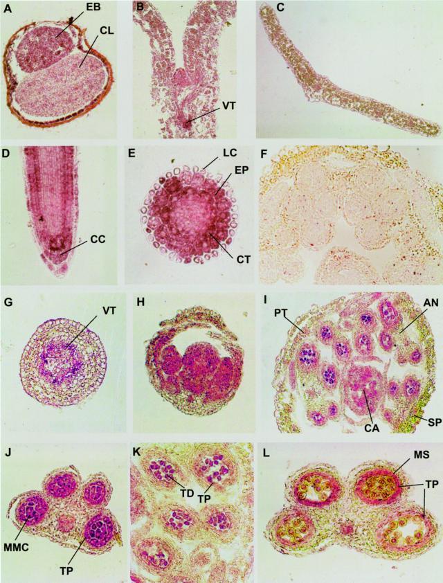
Figure 2.
Immunolicalization of Rop proteins in various Arabidopsis tissues. Ten-micrometer cryosections of Arabidopsis tissues were incubated with anti-Rop1Ps antibodies and alkaline phosphatase-conjugated secondary antibodies as described in text. A, Cross section of an imbibed seed. B, Longitudinal section of a shoot apex and cotyledons of seedling. C, Cross section of a rosette leaf. D, Longitudinal section of a root tip. E, Cross section of a root tip near the elongation zone of the root. F, Cross section of a closed flower bud stained with preimmune control. G, A cross section of a stem. H, Longitudinal section of a young flower bud. I, Cross section of a closed flower. J, K, and L, Cross sections of anthers at the microspore mother cell, tetrad, and early microspore stages, respectively. Purple color indicates Rop staining, whereas yellow and green colors indicate anthocyanin and chlorophyll pigments from sections of frozen tissues.

