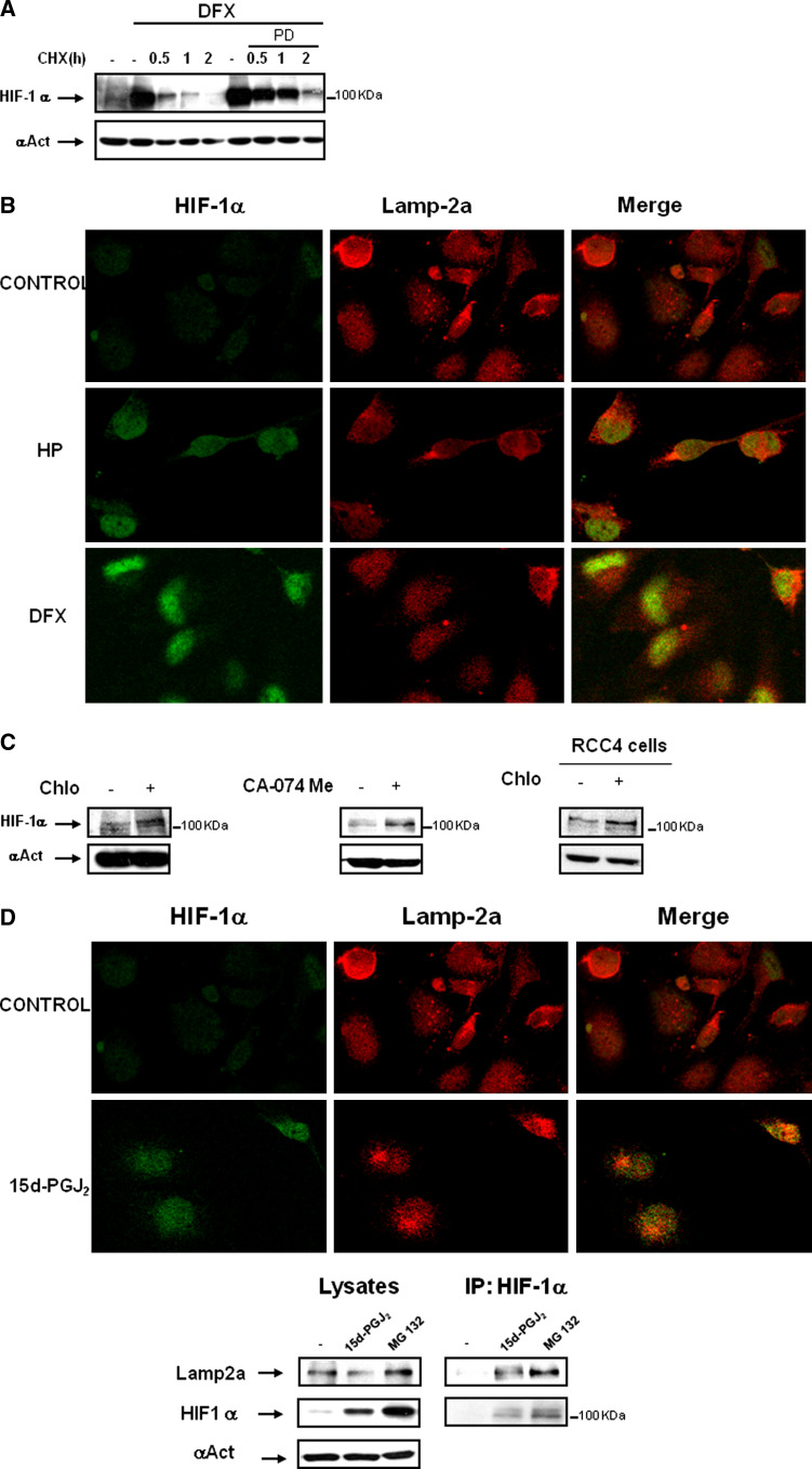
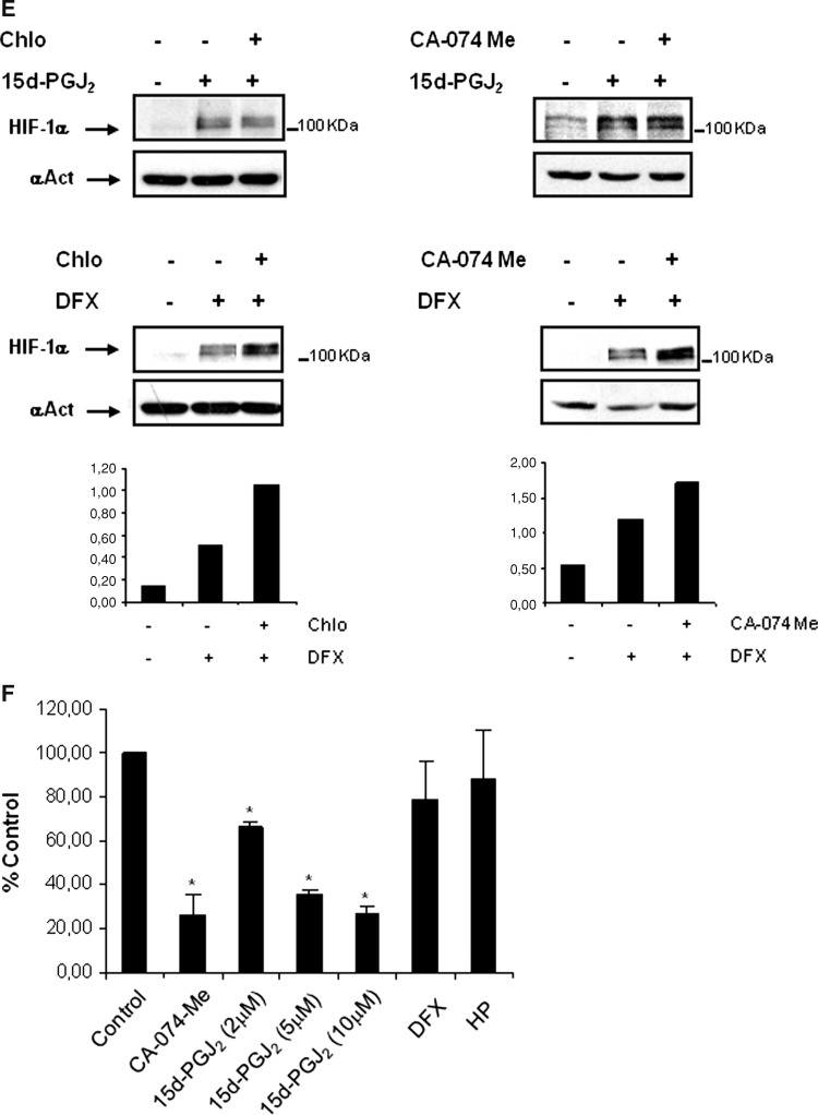
Fig. 4.
Effect of 15d-PGJ2 on lysosomal activity and its contribution to HIF-1α stabilization. a The effect of PD150606 calpain inhibitor on HIF-1α half-life was determined in HK-2 cells. Cells were incubated for 6 h with 380 μM DFX in the presence or absence of 50 μM PD150606. Thereafter, the protein translation inhibitor cycloheximide (CHX, 50 μg/ml) was added and incubation continued for up to 2 h. HIF-1α protein levels were determined by Western blot. b Immunofluorescence experiments were performed to determine whether HIF-1α, accumulated under several conditions, co-localizes with the lysosomal protein Lamp-2a. Role of lysosomes in HK-2 cells under hypoxia or DFX treated cells versus normoxic conditions. HK-2 cells were incubated for 6 h with 380 μM DFX or incubated under hypoxic conditions (1% O2). Then, cells were fixed and incubated with anti-HIF-1α and anti-Lamp-2a antibodies as described in “Materials and methods”. Immunofluorescence for Lamp-2a (red) and HIF-1α (green) is shown. c Accumulation of HIF-1α by lysosome inhibitors in normoxia. Cells were incubated for 24 h in the presence or absence of 50 μM chloroquine or 10 μM CA-074 Me. After incubation, cells were lysed, and HIF-1α levels were determined by Western blot analysis. d HK-2 cells were incubated for 6 h with 2 μM 15d-PGJ2. Then, cells were fixed and co-incubated with anti-HIF-1α and anti-Lamp-2a antibodies as described in “Materials and methods”. Immunofluorescence for Lamp-2a (red) and HIF-1α (green) is shown. As cells were incubated in parallel with DFX and hypoxia treatment, the control is the same as shown in Fig. 2b. Association of HIF-1α with lysosome marker Lamp-2a was assessed by immunoprecipitating the cells after 6 h in the presence of 15d-PGJ2 (2μM) or MG132 (10μM) as control of HIF-1α accumulation with an anti-HIF-1α antibody as described in “Materials and methods”. Immunoprecipitates were then probed by Western blot analysis using anti-HIF-1α or Lamp2a antibodies. Input controls show protein expression in cell lysates. A representative experiment out of three is shown. e Effect of chloroquine (Chlo) or the cathepsin B inhibitor CA-074 Me on 15d-PGJ2 or DFX-mediated HIF-1α stabilization. Cells were incubated for 24 h with 2 μM 15d-PGJ2 or 380 μM DFX in the presence or absence of 50 μM chloroquine or 10 μM CA-074 Me. HIF-1α expression levels were analyzed by Western Blot. f To determine the effect of 15d-PGJ2 on cathepsin B activity, cells were incubated for 24 h with 10 μM cathepsin B inhibitor CA-074Me, 2-5-10 μM 15d-PGJ2, 380 μM DFX or under hypoxic conditions (1% O2). Cathepsin B activity was measured as described in “Materials and methods”. The experiment was performed three times and each bar is the mean ± SD of the obtained results. *P < 0.01 versus control


