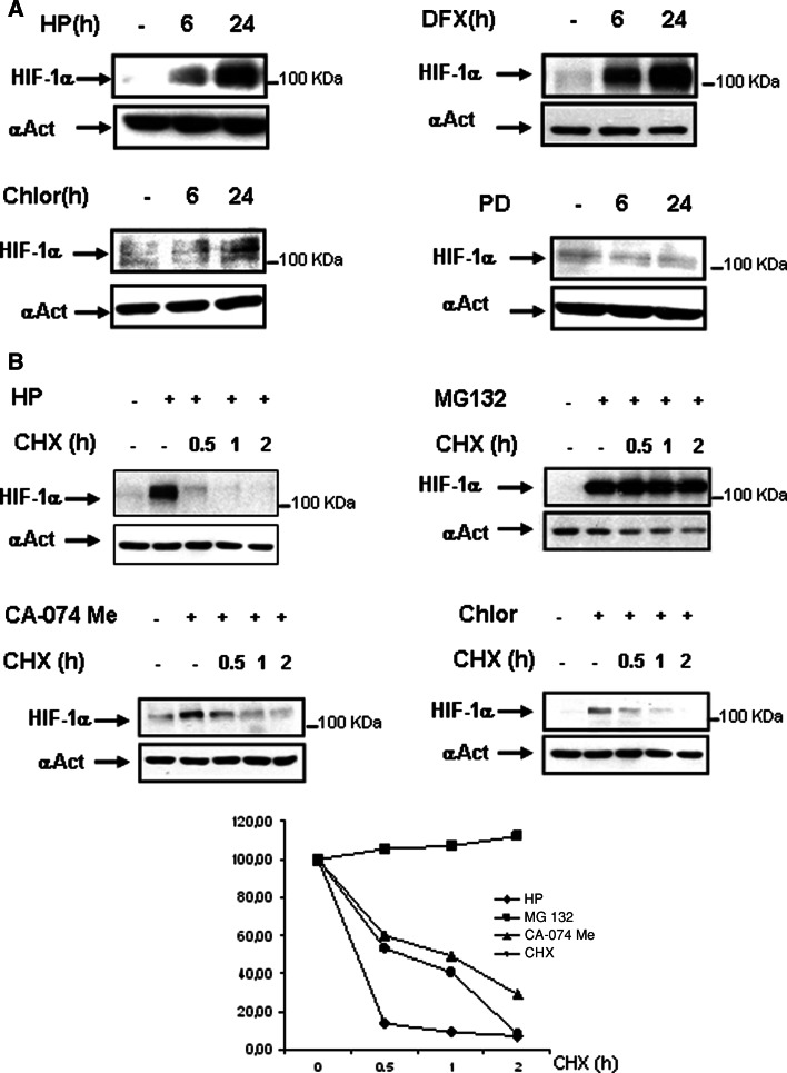
Fig. 5.
Comparative studies of the relative efficiency and kinetics of the three proposed pathways for HIF-degradation. a HK-2 cells were incubated for 6 or 24 h in the presence of 380 μM DFX; 50 μM calpain inhibitor PD150606 (PD), 50 μM clhoroquine or under hypoxia (1% O2). b Cells were incubated with 10 μM MG132 or under hypoxia (1% O2) for 6 h or with cloroquine (50 μM) or CA-074 Me (10 μM) for 24 h. Thereafter, 50 μg/ml of the protein translation inhibitor cycloheximide (CHX,) was added and incubation continued for up to 2 h. Cells were lysed immediately, and HIF-1α levels were determined by Western blot analysis. As a loading control, blots were reprobed with anti-α-actin. Each experiment was performed at least three times and a representative one is shown. Densitometric analysis is shown below

