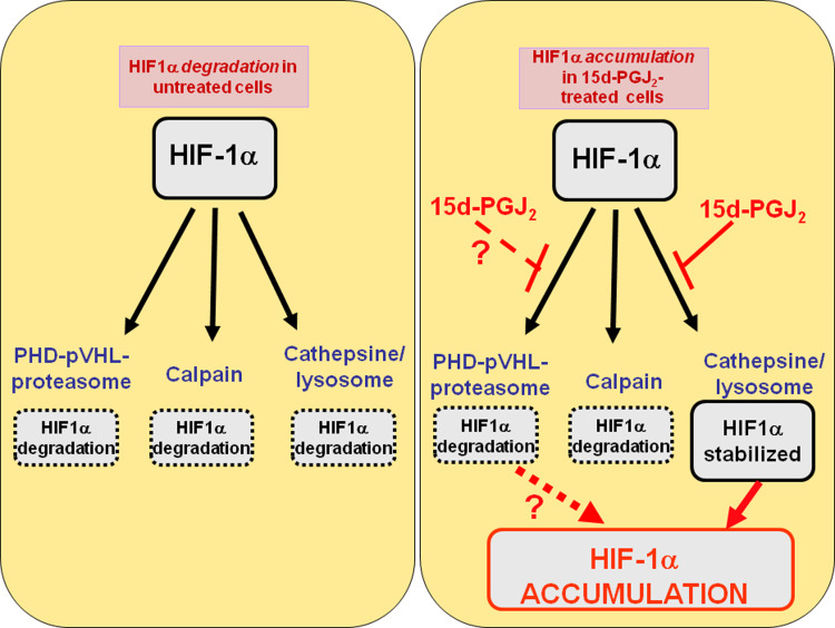
Fig. 6.
Enzymatic systems involved in the degradation of HIF-1α in human proximal tubular renal cells HK-2. Left: there are three operative enzymatic systems for the degradation of HIF-1α in HK-2 cells: proteasome (mainly through the PHD-pVHL-pathway, but also through other PHD-pVHL independent pathways such as this triggered by Hsp-90 inhibitor geldanamycin), calcium/calpain and cathepsin B/lysosome. Right: incubation with 15d-PGJ2 results in HIF-1α stabilization through the inhibition of cathepsin B/Lysosome, although the high degree of stabilization of HIF-1α strongly suggests that 15d-PGJ2 could also inhibit the PHD-pVHL-dependent proteasomal degradation of HIF-1α

