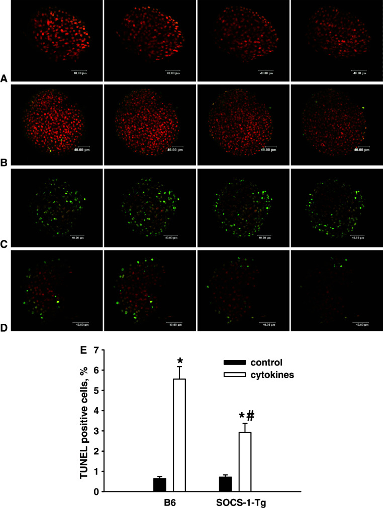
Fig. 2.
Expression of SOCS-1 protects islet cells from cytokine-induced cell death. B6 or SOCS-1-Tg mouse islets were incubated with or without the mixture of IL-1β, TNFα and IFNγ for 40 h. Confocal images of B6 control islet (a), SOCS-1-Tg control islet (b), B6 islet incubated with cytokines (c), SOCS-1-Tg islet incubated with cytokines (d), after nick translation labelling of DNA strand breaks (TUNEL) and double staining with FITC/PI. Apoptotic cells have a green colour. Bar width 40 μm. Several confocal images of each islet are represented. e Percentage of TUNEL positive cells detected by confocal microscopy. Black bars control islets; white bars cytokine-treated islets. Results are means ± SEM of 74–103 islets from five B6 and six SOCS-1-Tg mice. *P < 0.0005 relative to untreated control islets of the same genotype, # P < 0.05 relative to B6 islets treated with cytokines

