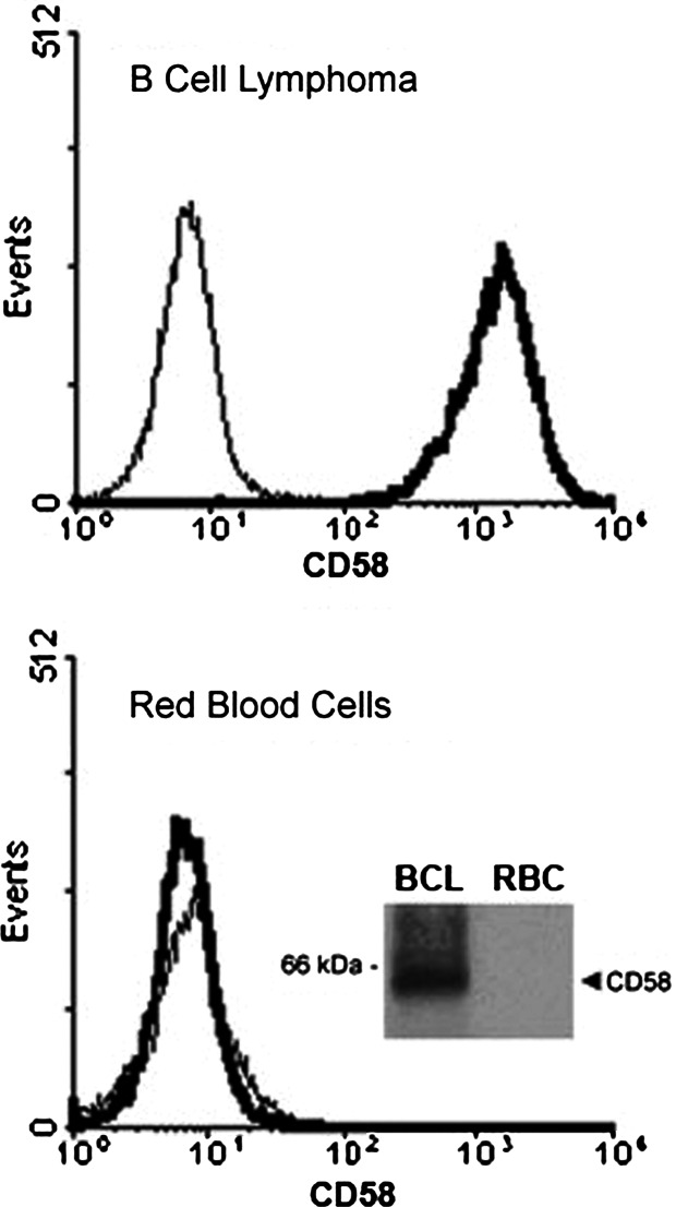
Fig. 3.
Human RBC lack expression of LFA-3/CD58. Red blood cells (RBC) and B cell lymphoma (BCL) cells were cell surface labeled with anti-LFA-3/CD58 antibodies (TS2/9) followed by RAM-FITC conjugated antibodies and acquired in a FACSCalibur. Histograms show CD58 expression (thick line) in BCL cells (upper histogram) and RBC (lower histogram). Background staining with RAM-FITC (thin line) is shown. Inset: BCL cells and RBC were surface biotinylated, lysed and LFA-3/CD58 immunoprecipitated with TS2/9 antibodies as described in “Materials and methods.” Aliquots of the immunoprecipitates were resolved in a SDS/PAGE, blotted and visualized by the ECL technique. The band corresponding to LFA-3/CD58 is indicated

