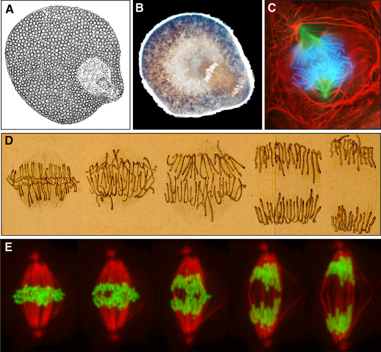
Fig. 1.
a First illustration of anaphase during the division of the first micromeres in the worm Rhynchelmis by A. Kowalevski (adapted from [2]). b Contemporary view of micromere formation in the sand dollar embryo (courtesy of George von Dassow, University of Washington, WA, USA). c Newt lung cell in anaphase as viewed by fluorescence microscopy (courtesy of Conly Rieder, Wadsworth Center, NY, USA). d Sequence of the anaphase movement in a plant cell as originally depicted by Strasburger (adapted from [4]). e Sequence of the anaphase movement from a time-lapse movie of a Drosophila S2 cell stably expressing mCherry-α-tubulin (red) and GFP-H2B-Histone (green) (courtesy of Sara Moutinho-Pereira, IBMC, University of Porto, Portugal). Note the simultaneity of anaphase A and B

