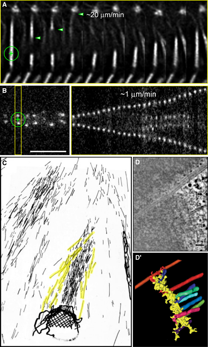
Fig. 4.
a Laser-mediated severing of a k-fiber in a Drosophila S2 cell stably expressing GFP-α-tubulin during metaphase. Note the fast depolymerization of the pole-proximal fragment and that the chromosome which remains attached to the severed k-fiber maintains its equatorial position (adapted from [52]). b Laser-mediated severing of the centromeric region in a Drosophila S2 cell stably expressing CID-GFP during metaphase. Note the slow poleward migration of each daughter kinetochore after surgery. Scale bar (a,b) 5 μm. c 3D-electron microscope reconstruction of a severed k-fiber from crane flies after irradiation with a UV microbeam (adapted from [177]). Non-kinetochore microtubules in the vicinity of the resulting k-fiber stub were pseudocolored in yellow. d Single slice from a tomographic reconstruction of a Ptk1 kinetochore showing both end-on and lateral MT binding (courtesy from Yimin Dong and Bruce McEwen, Wadsworth Center, NY, USA). Scale bar 100 nm. d’ 3D surface rendering of the 3D volume of the same kinetochore

