Fig. 2.
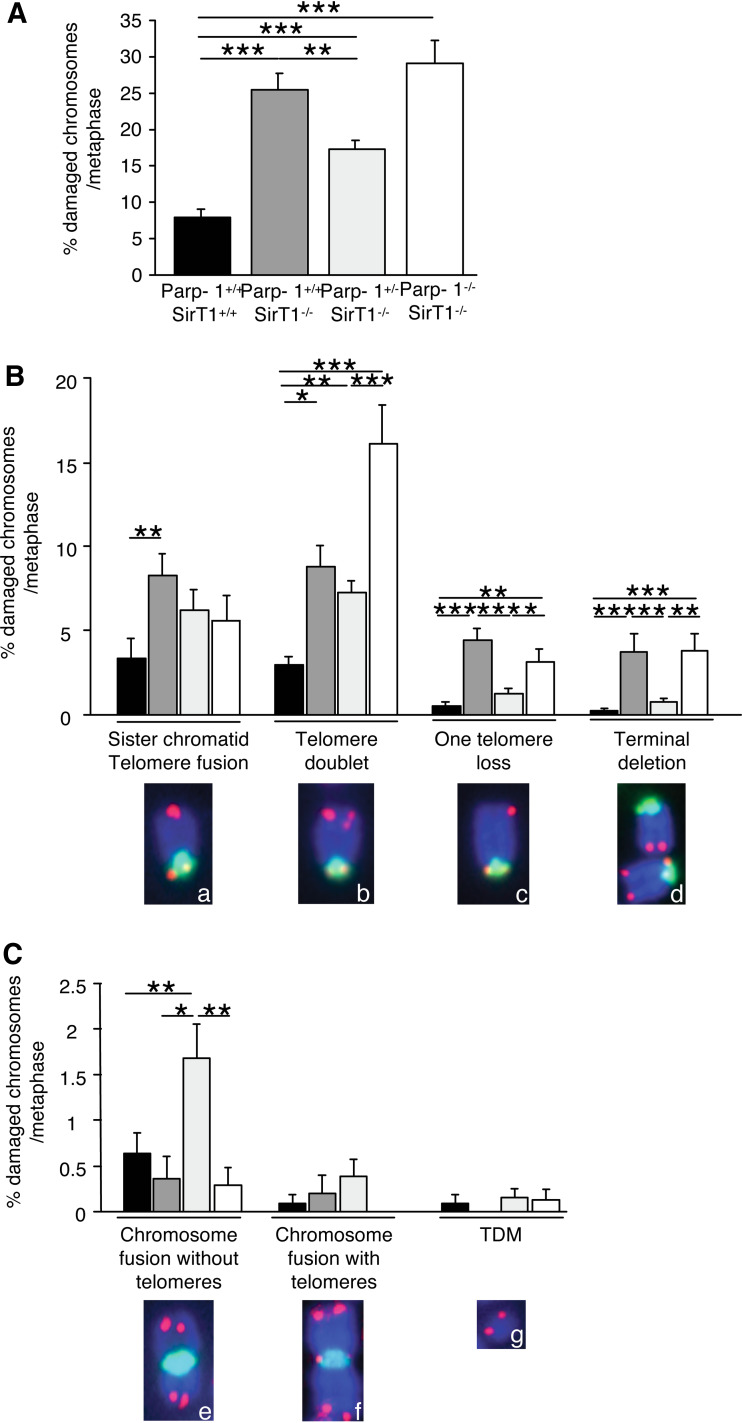
Telomere instability in Parp-1+/+;SirT1−/−, Parp-1+/−;SirT1−/− and Parp-1−/−;SirT1−/− 3T3 cell lines. Increased spontaneous telomere aberrations in Parp-1+/+;SirT1−/−, Parp-1+/−;SirT1−/− and Parp-1−/−;SirT1−/− 3T3 cells compared to Parp-1+/+;SirT1+/+ control cells. Telomere aberrations were detected by FISH on metaphase spreads as described in Materials and Methods. Total (A) or each type (B,C) of telomere aberrations are expressed as percentages of damaged chromosomes per metaphasis. The different types of telomere aberrations given in (B) are: chromosomes with sister chromatid telomere fusion (insert a), chromosomes with one extra-telomere signal referred to as telomere doublets (insert b), chromosomes with one telomere loss (insert c), chromosomes with terminal deletion (insert d), fused chromosomes without (insert e) or with telomeres (insert f) at the fusion points, and telomeric DNA-containing double minutes chromosomes (TDM, insert g). Inserts illustrate the different telomere aberrations identified in Parp-1+/+;SirT1−/− cells except for TDM identified in Parp-1−/−;SirT1−/− cells (green, pan-centromeric probe; red, PNA probe). Results were obtained from n = 18, 21, 20, and 15 metaphases for Parp-1+/+;SirT1+/+, Parp-1+/+;SirT1−/−, Parp-1+/−;SirT1−/−, and Parp-1−/−;SirT1−/− cells, respectively. Ordinary ANOVA tests revealed significant differences between genotypes intelomere aberrations (ANOVA, F 3,67 = 23.42, P < 0.0001) (A), telomere doublet (F 3,67 = 9.11, P < 0.0001), one telomere loss (F 3,67 = 13.29, P < 0.0001), terminal deletion (F 3,67 = 8.098, P < 0.001) (B), and chromosome fusion with telomeres (F 3,67 = 5.32, P = 0.024) (C). P value was calculated by Fischer’s test: P *< 0.05, **P < 0.01, ***P < 0.001

