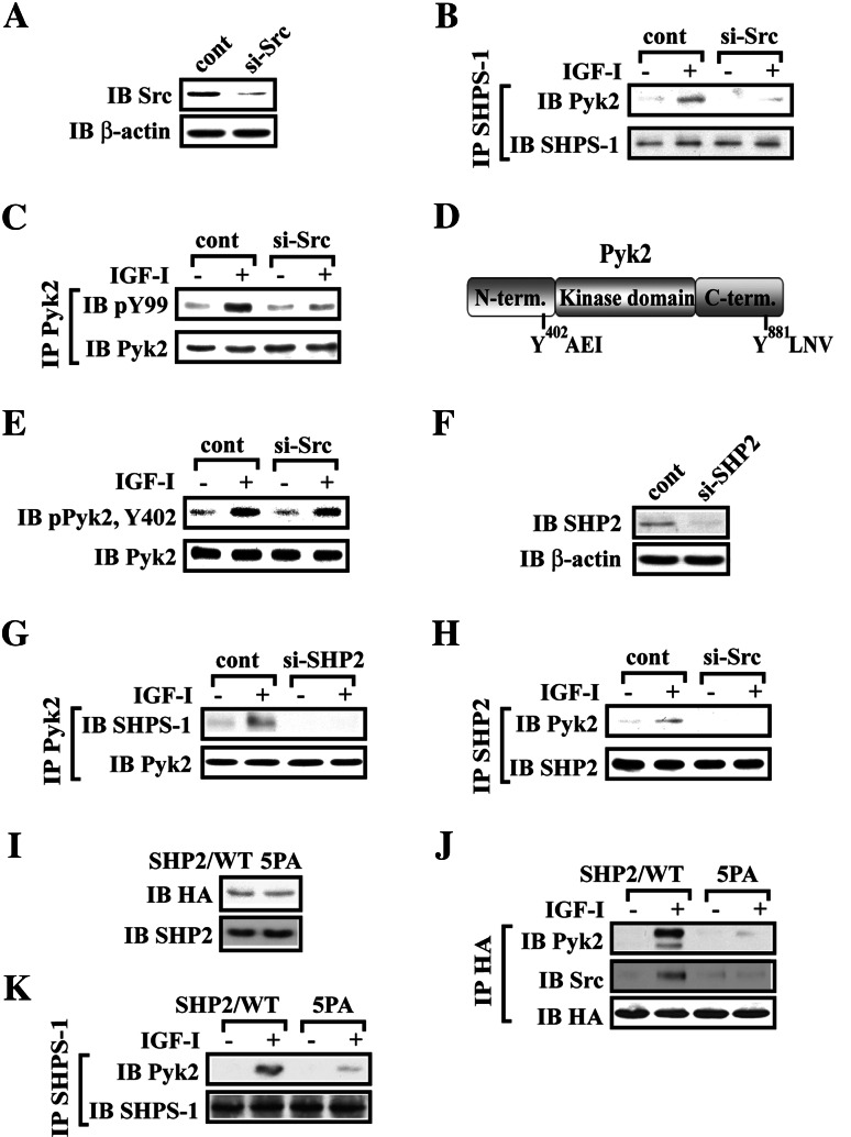
Fig. 2.
Src or SHP2 knock-down attenuates Pyk2 recruitment to SHPS-1. a SMCs were transduced with LacZ shRNA (control) or Src shRNA template plasmid and analyzed for Src protein expression. Cell lysates were immunoblotted with anti-Src antibody. The blot was stripped and reprobed with anti-β-actin antibody as a loading control. Quiescent Src knock-down SMCs were stimulated with IGF-I. Cell lysates were immunoprecipitated (IP) with anti-SHPS-1 antibody (b) or anti-Pyk2 antibody (c) followed by immunoblotting with the antibody indicated (upper panel). To control for loading, the blot was stripped and reprobed with the antibody indicated (lower panel). d Diagram of the pyk2 phosporylation sites. The autophosphorylation of Pyk2 at Try402 provides a binding site for Src tyrosine kinase recruitment, and subsequently, Src phosphorylates Pyk2 on Tyr881 which provides a binding site for Grb2. e Autophosphorylation of Pyk2 at Tyr402 in Src knock-down cells. Twenty micrograms of cell lysate was directly immunoblotted for phospho-Pyk2 (Y402). The blot was stripped and reprobed with anti-Pyk2 antibody as a loading control. f SMCs were transduced with LacZ shRNA (control) or SHP2 shRNA template plasmid and analyzed for SHP2 protein expression. Cell lysates were immunoblotted with anti-SHP2 antibody. The blot was stripped and reprobed with anti-β-actin antibody as a loading control. Quiescent SHP2 knock-down or Src knock-down SMCs were stimulated with IGF-I. Cell lysates were immunoprecipitated (IP) with anti-Pyk2 antibody (g) or anti-SHP2 antibody (h) followed by immunoblotting with the antibody indicated (upper panel). To control for loading, the blot was stripped and reprobed with the antibody indicated (lower panel). i SMCs expressing SHP2/WT or SHP2/5PA were lysed. The blot was probed with an anti-HA antibody or anti-SHP2 antibody to detect expression of SHP2/WT or SHP2/5PA. j Quiescent SHP2/WT- or SHP2/5PA-expressing SMCs were stimulated with IGF-I for 5 min. Cell lysates were immunoprecipitated (IP) with anti-HA antibody and immunoblotted (IB) for the protein of interest. To control the loading, the blots were stripped and reprobed with anti-HA antibody. k Quiescent SHP2/WT or SHP2/5PA expressing SMCs were stimulated with IGF-I for 5 min. Cell lysates were immunoprecipitated (IP) with anti-SHPS-1 antibody and immunoblotted (IB) for Pyk2. To control the loading, the blots were stripped and reprobed with anti-SHPS-1 antibody

