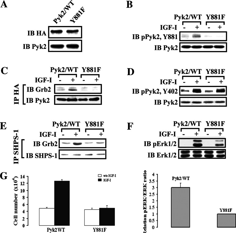
Fig. 6.
IGF-I-stimulated Pyk2 Tyr881 phosphorylation is essential for Grb2 recruitment to SHPS-1 and leads to Erk1/2 phosphorylation and cell proliferation. a SMCs expressing Pyk2/WT or Pyk2/Y881F were lysed and the lysates immunoblotted for HA or Pyk2 to detect expression of Pyk2/WT or Pyk2/Y881F. b–e Quiescent Pyk2/WT- or Pyk2/Y881F-expressing SMCs were stimulated with IGF-I for 2 or 5 min. Cell lysates were immunoblotted for detection of phospho-Pyk2 (Y881) (b) or phospho-Pyk2 (Y402) (d). The blots were stripped and reprobed with anti-Pyk2 antibody as a loading control. Cell lysates from the same experiment were immunoprecipitated (IP) with anti-HA antibody (c) or anti-SHPS-1 antibody (e) and immunoblotted (IB) with anti-Grb2 antibody. To control the loading, the blots were stripped and reprobed with the antibody indicated (lower panels). f Twenty micrograms of cell lysate from the same experiment was used for detection of phospho-Erk1/2. The blots were stripped and reprobed with anti-Erk1/2 antibody as a loading control. The protein levels were quantified using scanning densitometry. The graph shows the mean result from three independent experiments expressed as relative pERK/ERK ratio that was calculated from arbitrary scanning units (f, lower panel). g Proliferation of Pyk2/WT or Pyk2/Y881F mutant cells following IGF-I stimulation. Cell proliferation was determined as described in “Materials and methods”. The results represent a mean value (±SE) of six independent experiments

